. 2022 Feb 17:13:828263.
doi: 10.3389/fimmu.2022.828263. eCollection 2022.

Development of a Dendritic Cell/Tumor Cell Fusion Cell Membrane Nano-Vaccine for the Treatment of Ovarian Cancer

Affiliations

Development of a Dendritic Cell/Tumor Cell Fusion Cell Membrane Nano-Vaccine for the Treatment of Ovarian Cancer

Lei Zhang et al. Front Immunol. .

Abstract

Ovarian cancer (OC) is a malignant tumor that seriously affects women's health. In recent years, immunotherapy has shown great potential in tumor treatment. As a major contributor of immunotherapy, dendritic cells (DCs) - based tumor vaccine has been demonstrated to have a positive effect in inducing immune responses in animal experiments. However, the effect of tumor vaccines in clinical trials is not ideal. Therefore, it is urgent to improve the existing tumor vaccines for tumor treatment. Here, we developed a fusion cell membrane (FCM) nano-vaccine FCM-NPs, which is prepared by fusing DCs and OC cells and coating the FCM on the poly (lactic-co-glycolic acid) (PLGA) nanoparticles (NPs) loaded with the immune adjuvant CpG-oligodeoxynucleotide (CpG-ODN). The fusion process promoted the maturation of DCs, thus up-regulating the expression of costimulatory molecule CD80/CD86 and accelerating lymph node homing of DCs. Furthermore, FCM-NPs has both the immunogenicity of tumor cells and the antigen presenting ability of DCs, it can stimulate naive T lymphocytes to produce a large number of tumor-specific cytotoxic CD8+ T lymphocytes. FCM-NPs exhibited strong immuno-activating effect both in vitro and in vivo. By establishing subcutaneous transplanted tumor model, patient-derived xenograft tumor model and abdominal metastatic tumor model, FCM-NPs was proved to have the effect of delaying the growth and inhibiting the metastasis of OC. FCM-NPs is expected to become a new tumor vaccine for the treatment of ovarian cancer.

Keywords: CpG-ODN; cytotoxic T lymphocytes; dendritic cell; fusion cell membrane; ovarian cancer.


Conflict of interest statement

The authors declare that the research was conducted in the absence of any commercial or financial relationships that could be construed as a potential conflict of interest.

Figures

Figure 1
Figure 1
Schematic illustration for the preparation of FCM-NPs. Fusion cells were first prepared from ovarian cancer (OC) cells and mouse dendritic cells (DCs) by PEG method. FCs membrane was then extracted and mixed with CpG ODN-loaded PLGA-NPs, and the FCM-NPs was prepared by extrusion method.
Figure 2
Figure 2
Identification and characterization of FCM-NPs. (A) The fusion of DCs and OC cells was observed through CLSM, red fluorescence of PKH26-labelled ID8 cells and green fluorescence of CFSE-labelled DCs. Scale bar = 200 μm. (B) Flow cytometry analyses of the CD133-PE on ID8 cells, MHC II-FITC on DCs and the double-labeling on FCs. (C) SDS-PAGE analysis of the proteins of ID8 cell membrane (CCM), dendritic cell membrane (DCM) and fusion cell membrane (FCM). (D) Western blot analysis of the protein biomarkers of cell membrane, cytoplasm and nucleus in FC and FCM. (E) TEM images of FCM-NPs. Scale bar = 100 nm. (F) ζ-potential of the NPs, CCM-NPs, DCM-NPs and FCM-NPs.
Figure 3
Figure 3
In vitro immune activation of FCM-NPs. (A) Flow cytometry analyses of the mature biomarkers CD86-APC and CD80-PE of BMDCs co-incubated with NPs, CCM-NPs, DCM-NPs and FCM-NPs. (B) Maturation rate of BMDCs in each group. (C) Levels of TNF-α and IL-6 secreted by BMDCs determined by ELISA. (D) Flow cytometry analyses of the expression of CD8 and CD4 in splenic lymphocytes co-incubated with BMDCs activated by NPs, CCM-NPs, DCM-NPs and FCM-NPs. *P < 0.05, **P < 0.01, ***P < 0.001 vs NPs group.
Figure 4
Figure 4
In vivo immune activation of FCM-NPs. (A) In vivo fluorescence imaging of mice at different time points after vaccine injection. (B) Fluorescence imaging of spleen and lymph node of each group of mice 36 h after vaccine immunization. (C) The secretion of inflammatory cytokines TNF-α and IL-6 in serum detected by ELISA. **P < 0.01, ***P < 0.001 vs PBS group, ## P < 0.01, ### P < 0.001 vs NPs group. (D) Flow cytometry analyses of the expression of CD3 and CD8 in spleen of mice treated with PBS, NPs, CCM-NPs, DCM-NPs and FCM-NPs. (E) Flow cytometry analyses of IFN-γ+ and CD8+ effector T cell in each group. (F) Flow cytometry analyses of the CD4+ CD25+ Foxp3+ Treg cells. *P < 0.05, **P < 0.01, ***P < 0.001 vs PBS group.
Figure 5
Figure 5
Immune stimulation and therapeutic effect of FCM-NPs on subcutaneous transplanted tumor. (A) Schematic diagram of modeling and treatment of subcutaneous transplanted tumor in mice. (B) Changes in tumor size of mice in PBS, NPs, CCM-NPs, DCM-NPs and FCM-NPs groups. *P < 0.05, **P < 0.01, ***P < 0.001 vs PBS group. (C) Images of tumors after the mice were sacrificed. (D) Immunofluorescence staining of CD3 and CD8 in tumor sections. Scale bar = 200 μm.
Figure 6
Figure 6
Immune stimulation and therapeutic effect of FCM-NPs on PDX tumor model. (A) Schematic diagram of modeling and treatment of PDX tumor in mice. (B) The size changes of P1DX and P2DX tumor in PBS, NPs, CCM-NPs, DCM-NPs and FCM-NPs groups. (C) In vivo fluorescence detection of the vaccine selectivity in double tumor mode. Scale bar = 200 μm. (D) Immunofluorescence staining of CD3 and CD8 in P1DX and P2DX tumor sections.
Figure 7
Figure 7
Therapeutic effect of FCM-NPs on abdominal metastatic ovarian cancer model. (A) Schematic diagram of modeling and treatment of abdominal metastatic tumor in mice. (B) Images of adnexa uteri of mice treated with PBS, NPs, CCM-NPs, DCM-NPs and FCM-NPs. (C) Weight of adnexa uteri in mice of each group. *P < 0.05, **P < 0.01 vs PBS group. (D) The quantitative results of the metastatic tumor nodules on the abdominal wall. **P < 0.01, ***P < 0.001 vs PBS group.
Figure 8
Figure 8
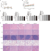
Biosafety evaluation of FCM-NPs. (A) The cytotoxicity of FCM-NPs to hepatocyte HL7702 and renal tubular epithelial cell HK-2 was assessed by MTT assay. (B) Body weight changes of mice with subcutaneous transplanted tumor and vaccines treatment. (C) Liver and kidney function indexes. (D) HE staining images of major organs of subcutaneous transplanted tumor model mice after vaccine treatment.

References

    1. Cornelison R, Llaneza DC, Landen CN. Emerging Therapeutics to Overcome Chemoresistance in Epithelial Ovarian Cancer: A Mini-Review. Int J Mol Sci (2017) 18:2171. doi: 10.3390/ijms18102171 - DOI - PMC - PubMed
    1. Pokhriyal R, Hariprasad R, Kumar L, Hariprasad G. Chemotherapy Resistance in Advanced Ovarian Cancer Patients. biomark Cancer (2019) 11:1179299x19860815. doi: 10.1177/1179299X19860815 - DOI - PMC - PubMed
    1. Ostrand-Rosenberg S. Tumor Immunotherapy: The Tumor Cell as an Antigen-Presenting Cell. Curr Opin Immunol (1994) 6:722–7. doi: 10.1016/0952-7915(94)90075-2 - DOI - PubMed
    1. Yang Y. Cancer Immunotherapy: Harnessing the Immune System to Battle Cancer. J Clin Invest (2015) 125:3335–7. doi: 10.1172/JCI83871 - DOI - PMC - PubMed
    1. Alard E, Butnariu AB, Grillo M, Kirkham C, Zinovkin DA, Newnham L, et al. . Advances in Anti-Cancer Immunotherapy: Car-T Cell, Checkpoint Inhibitors, Dendritic Cell Vaccines, and Oncolytic Viruses, and Emerging Cellular and Molecular Targets. Cancers (Basel) (2020) 12(7):1826. doi: 10.3390/cancers12071826 - DOI - PMC - PubMed

Publication types

Substances