. 2022 Feb 17:13:835348.
doi: 10.3389/fimmu.2022.835348. eCollection 2022.

LPS Guides Distinct Patterns of Training and Tolerance in Mast Cells

Affiliations

LPS Guides Distinct Patterns of Training and Tolerance in Mast Cells

Marco De Zuani et al. Front Immunol. .

Abstract

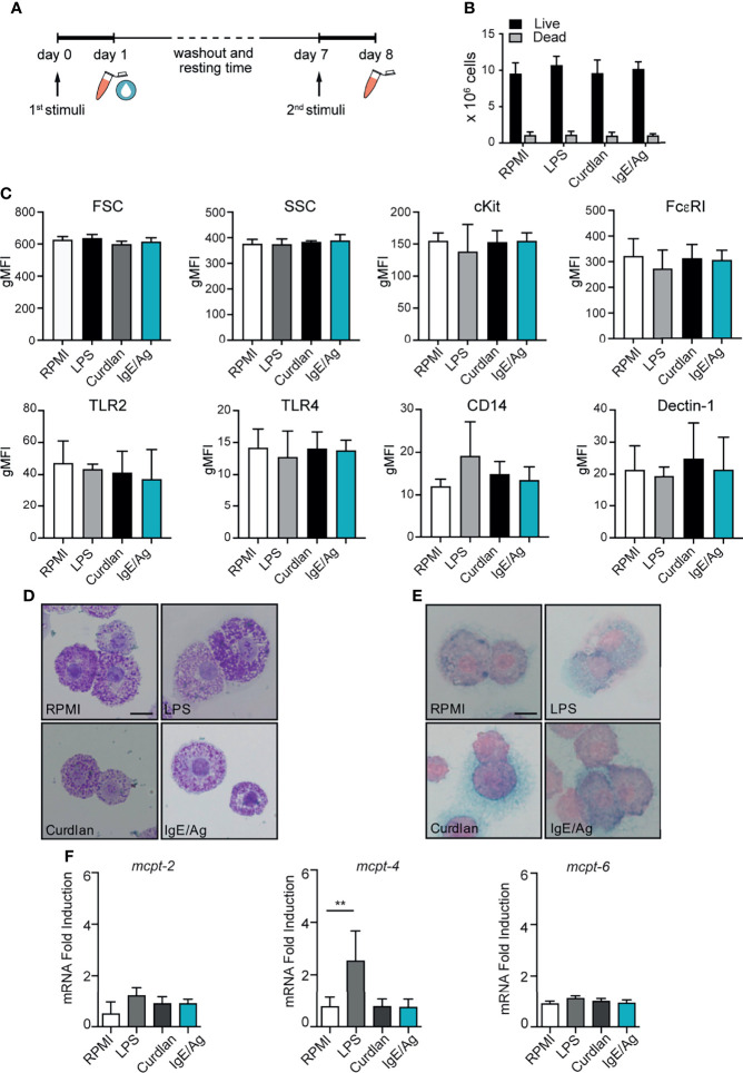
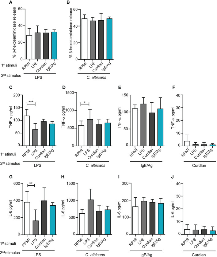
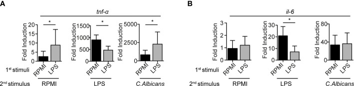
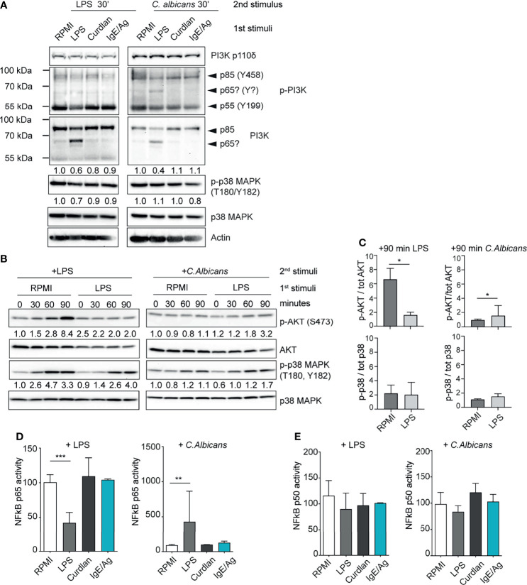
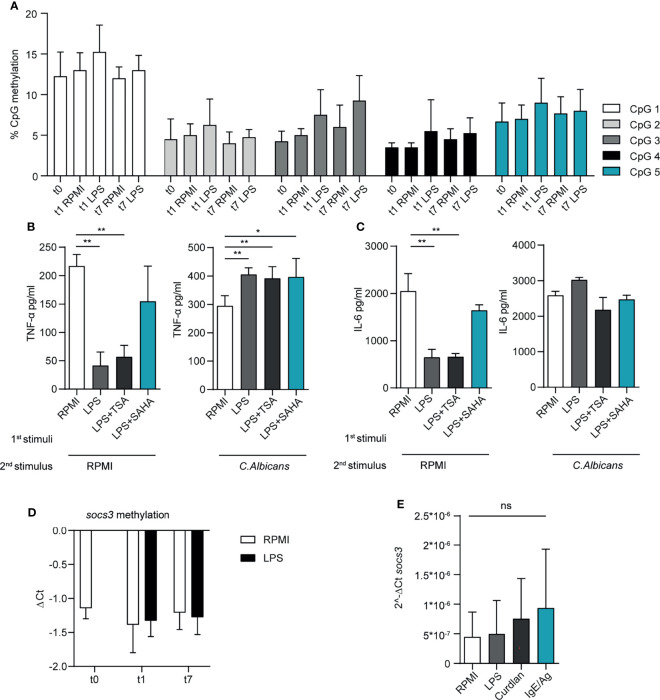
Mast cells (MCs) are tissue-resident, long lived innate immune cells with important effector and immunomodulatory functions. They are equipped with an eclectic variety of receptors that enable them to sense multiple stimuli and to generate specific responses according on the type, strength and duration of the stimulation. Several studies demonstrated that myeloid cells can retain immunological memory of their encounters - a process termed 'trained immunity' or 'innate immune memory'. As MCs are among the one of first cells to come into contact with the external environment, it is possible that such mechanisms of innate immune memory might help shaping their phenotype and effector functions; however, studies on this aspect of MC biology are still scarce. In this manuscript, we investigated the ability of MCs primed with different stimuli to respond to a second stimulation with the same or different ligands, and determined the molecular and epigenetic drivers of these responses. Our results showed that, while the stimulation with IgE and β-glucan failed to induce either tolerant or trained phenotypes, LPS conditioning was able to induce a profound and long-lasting remodeling of the signaling pathways involved in the response against LPS or fungal pathogens. On one side, LPS induced a strong state of unresponsiveness to secondary LPS stimulation due to the impairment of the PI3K-AKT signaling pathway, which resulted in the reduced activation of NF-κB and the decreased release of TNF-α and IL-6, compared to naïve MCs. On the other side, LPS primed MCs showed an increased release of TNF-α upon fungal infection with live Candida albicans, thus suggesting a dual role of LPS in inducing both tolerance and training phenotypes depending on the secondary challenge. Interestingly, the inhibition of HDAC during LPS stimulation partially restored the response of LPS-primed MCs to a secondary challenge with LPS, but failed to revert the increased cytokine production of these cells in response to C. albicans. These data indicate that MCs, as other innate immune cells, can develop innate immune memory, and that different stimulatory environments can shape and direct MC specific responses towards the dampening or the propagation of the local inflammatory response.

Keywords: Candida albicans; LPS; cytokines; endotoxin tolerance; mast cell; trained immunity.


Conflict of interest statement

The authors declare that the research was conducted in the absence of any commercial or financial relationships that could be construed as a potential conflict of interest.

Figures

Figure 1
Figure 1
MC vitality and phenotype after the conditioning period. (A) Schematic representation of experimental protocol. BMMC were first stimulated for 24h with LPS, curdlan, IgE/Ag or left untreated (RPMI). Then cells were left to recover for 6 days, countedand tested for viability by means of trypan blue exclusion (B), analyzed for surface marker expression (C), stained with Toluidin blue (D) and Alcian-blu safranin (E) and analyzed for connective and mucosal proteases mRNA expression (F). Data are expressed as mean +SD from n>3 experiments; statistical analysis were performed with one-way Anova with Dunnet correction (**p < 0,01).
Figure 2
Figure 2
Effect of different priming on MC degranulation and cytokine release response to a secondary challenge. Degranulation of BMMCs primed with LPS (1mg/ml), curdlan (100mg/ml), IgE/Ag or RPMI was determined 6 days after cell recovery as percentages of released β-hexosaminidase in response to 30 min of stimulation with IgE/Ag (A) or ionomycin (B). BMMC primed with RPMI, LPS (1mg/ml), curdlan (100mg/ml) or IgE/Ag were subjected to a second stimulation with LPS (C–G), live C. Albicans (D–H), IgE/Ag (F–I) or curdlan (E–J). TNF-α (C–F) and IL-6 (G–J) levels were detected in culture supernatants after 24 hours. Data are expressed as mean +SD from n>4 experiments; statistical analysis were performed with one-way Anova with Dunnet correction (*p < 0,05 **p < 0,01; ***p < 0,001).
Figure 3
Figure 3
mRNA cytokine expression levels are impaired in LPS-primed BMMCs. mRNA expression of TNF- α (A) and IL-6 (B) were measured by rt-PCR on LPS-primed BMMC after 3 hours of stimulation with LPS (1mg/ml), live C. Albicans (MOI=1) or RPMI. Data are expressed as mean +SD from n=3 experiments. Statistical analysis were performed with paired Student t-test (*p < 0,05).
Figure 4
Figure 4
LPS-primed MCs differently activate the PI3K-AKT pathway. (A) Western Blot (WB) analysis of PI3K subunits and of phosphorylated p38 in differently primed-BMMC restimulated for 30 minutes with LPS or live C. Albicans (MOI=1). Numbers indicate the results of densitometric analyses calculated over endogenous protein expression. (B) Time-course WB analysis of phosphor Akt and phospho p38 in LPS primed-BMMC restimulated with LPS or live C. Albicans (MOI=1). Numbers indicate the results of densitometric analyses calculated over endogenous protein expression normalized versus t=0. (C) Mean + SD of densitometric analysis of n=3 independent experiments are reported. Statistical analyses were performed with paired Student t-test (*=p<0,05). NF-kB p65 (D) and p50 (E) activity in LPS primed BMMC was determined after 30 minutes form addition of the second stimuli. Results were expressed as fold percentages over control (RPMI). Mean + SD of n=3 independent experiments are reported. Statistical analysis were performed with one-way Anova with Dunnet correction (*p < 0,05 **p < 0,01; ***p < 0,001).
Figure 5
Figure 5
LPS priming affects the epigenetic landscape in MCs. (A) CpG methylation at TNF-α promoter region. A total of five CpG sites were analysed in the selected region, their level of methylation was determined by pyrosequencing. Mean + SD of n=3-5 independent experiments are reported. BMMC were primed with LPS (1μg/ml) in presence of HDAC inhibitors (SAHA 1nM; TSA 10nM), then washed, re-stimulated and analysed for TNF-α (B) and IL-6 (C) secretion upon second stimulation with LPS or C. albicans. Mean + SD of n=3 independent experiments are reported. (D) SOCS3 promoter CpG methylation was determined by real-time MSP. Methylation was calculated as difference among Ct values of unmethylated and methylated region. Mean + SD of n=2-6 independent experiments are reported. (E) SOCS3 mRNA expression in BMMC at day 7, i.e. 6 days after the first stimulation with LPS, curdlan and IgE/Ag. Mean + SD of n=3 independent experiments are reported. Statistical analysis were performed with one-way Anova with Dunnet correction (*p < 0,05 **p < 0,01; ***p < 0,001 ns, not significant).
Figure 6
Figure 6
Effect of repetitive LPS stimulation on BMMCs. Schematic representation of experimental protocol: 4-weeks old BMMCs were treated for 24h with LPS once a week for three weeks (A). Surface marker expression (B) and MCPT-4 mRNA expression (C) were evaluated at indicated times. The levels of TNF-α (D) and IL-6 (E) released by BMMC after the first, the second and the third dose of LPS are shown. The levels of TNF-α (F) and IL-6 (G) released by BMMC treated with one or more doses of LPS have been compared. Data are means +SD. Statistical analysis were performed with one-way Anova with Dunnet correction (*p < 0,05 **p < 0,01; ***p < 0,001).

References

    1. Netea MG, Domínguez-Andrés J, Barreiro LB, Chavakis T, Divangahi M, Fuchs E, et al. . Defining Trained Immunity and Its Role in Health and Disease. Nat Rev Immunol (2020) 20(6):375–88. doi: 10.1038/s41577-020-0285-6 - DOI - PMC - PubMed
    1. Netea MG, van der Meer JW. Trained Immunity: An Ancient Way of Remembering. Cell Host Microbe (2017) 21(3):297–300. doi: 10.1016/j.chom.2017.02.003 - DOI - PubMed
    1. Saeed S, Quintin J, Kerstens HH, Rao NA, Aghajanirefah A, Matarese F, et al. . Epigenetic Programming of Monocyte-to-Macrophage Differentiation and Trained Innate Immunity. Science (2014) 345(6204):1251086. doi: 10.1126/science.1251086 - DOI - PMC - PubMed
    1. Quintin J, Cheng SC, van der Meer JWM, Netea MG. Innate Immune Memory: Towards a Better Understanding of Host Defense Mechanisms. Curr Opin Immunol (2014) 29(1):1–7. doi: 10.1016/j.coi.2014.02.006 - DOI - PubMed
    1. Ifrim CD, Quintin J, Joosten L, Jacobs C, Jansen T, Jacobs L, et al. . Mihai G Netea. Trained Immunity or Tolerance: Opposing Functional Programs Induced in Human Monocytes After Engagement of Various Pattern Recognition Receptors. Clin Vaccine Immunol (2014) 21(4):534–45. doi: 10.1128/CVI.00688-13 - DOI - PMC - PubMed

Publication types

MeSH terms