. 2022 Feb 23:2022:2760147.
doi: 10.1155/2022/2760147. eCollection 2022.

Intravitreal Administration Effect of Adipose-Derived Mesenchymal Stromal Cells Combined with Anti-VEGF Nanocarriers, in a Pharmaceutically Induced Animal Model of Retinal Vein Occlusion

Affiliations

Intravitreal Administration Effect of Adipose-Derived Mesenchymal Stromal Cells Combined with Anti-VEGF Nanocarriers, in a Pharmaceutically Induced Animal Model of Retinal Vein Occlusion

Eleni Gounari et al. Stem Cells Int. .

Abstract

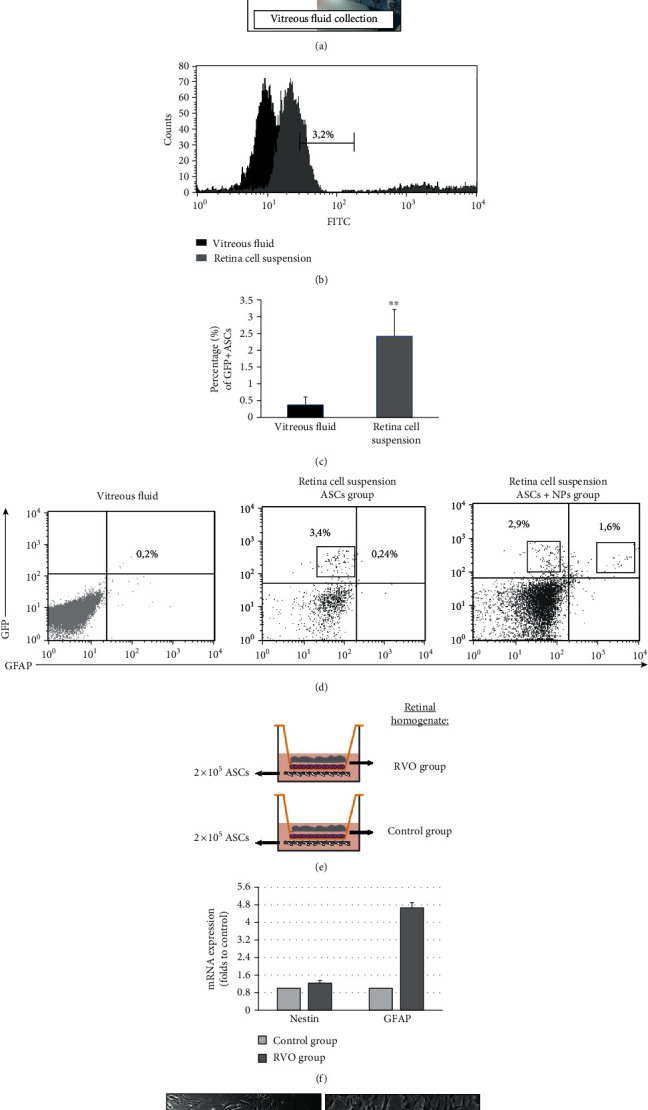
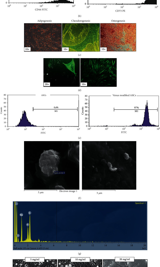
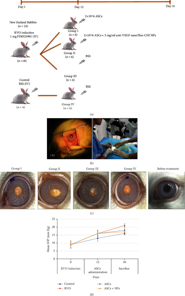
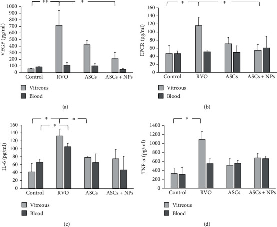
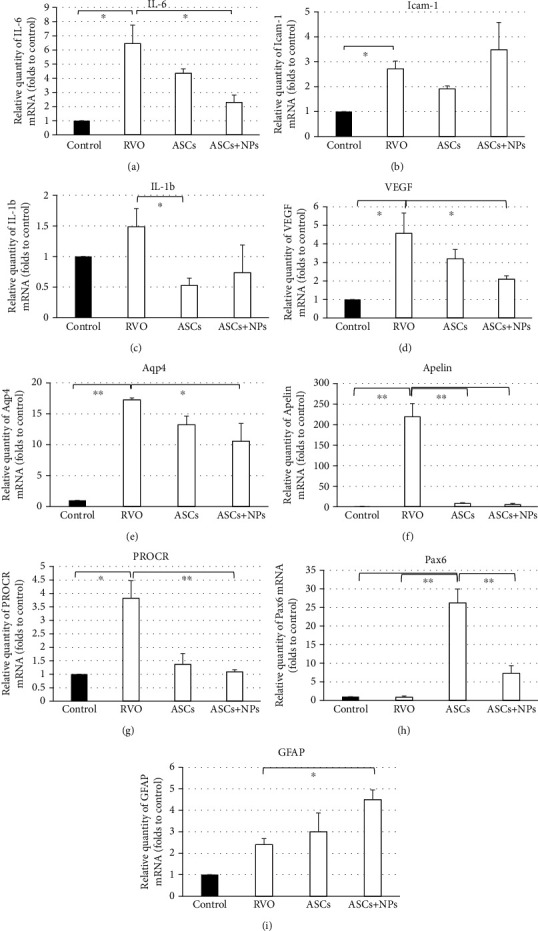
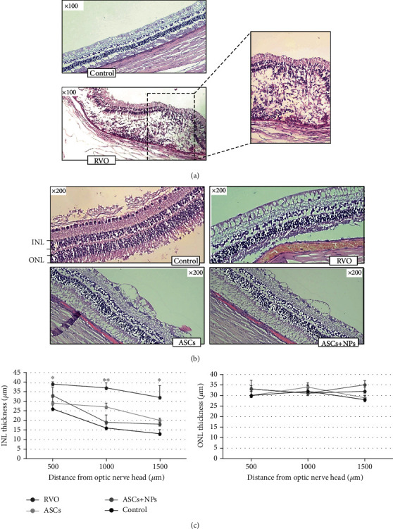
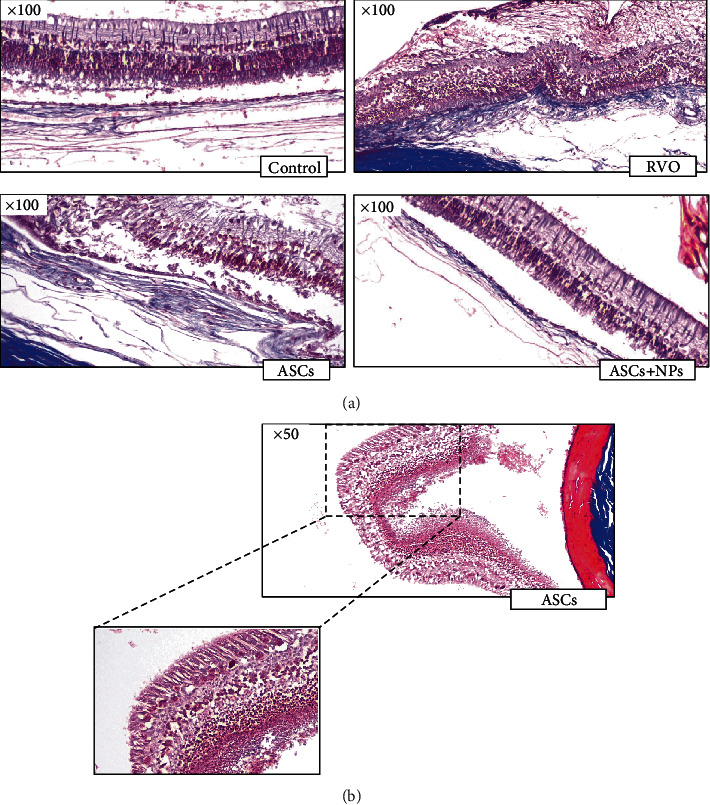
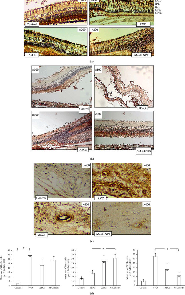
Antiangiogenic therapeutic agents (anti-VEGF) have contributed to the treatment of retinal vein occlusion (RVO) while mesenchymal stromal cell- (MSCs-) mediated therapies limit eye degeneration. The aim of the present study is to determine the effect of adipose-derived MSCs (ASCs) combination with nanocarriers of anti-VEGF in a pharmaceutically induced animal model of RVO. Nanoparticles (NPs) of thiolated chitosan (ThioCHI) with encapsulated anti-VEGF antibody were prepared. ASCs were isolated and genetically modified to secrete the green fluorescence GFP. Twenty-four New Zealand rabbits were divided into the I-IV equal following groups: ASCs, ASCs + nanoThioCHI-anti-VEGF, RVO, and control. For the RVO induction, groups I-III received intravitreal (iv) injections of MEK kinase inhibitor, PD0325901. Twelve days later, therapeutic regiments were administered at groups I-II while groups III-IV received BSS. Two weeks later, the retinal damage evaluated via detailed ophthalmic examinations, histological analysis of fixed retinal sections, ELISA for secreted cytokines in peripheral blood or vitreous fluid, and Q-PCR for the expression of related to the occlusion and inflammatory genes. Mild retinal edema and hemorrhages, limited retinal detachment, and vasculature attenuation were observed in groups I and II compared with the pathological symptoms of group III which presented a totally disorganized retinal structure, following of positive immunostaining for neovascularization and related to RVO markers. Important reduction of the high secreted levels of inflammatory cytokines was quantified in groups I and II vitreous fluid, while the expression of the RVO-related and inflammatory genes has been significantly decreased especially in group II. GFP+ ASCs, capable of being differentiated towards neural progenitors, detected in dissociated retina tissues of group II presenting their attachment to damaged area. Conclusively, a stem cell-based therapy for RVO is proposed, accompanied by sustained release of anti-VEGF, in order to combine the paracrine action of ASCs and the progressive reduction of neovascularization.


Conflict of interest statement

The authors declare that they have no conflict of interest.

Figures

Figure 1
Figure 1
Detection of administered GFP+ ASCs in dissociated retina tissues and in vitro differentiation capacity of ASCs towards neural cells as a response to specific tissue stimuli in culture. (a) The muscles and connective tissues of the rabbit eyes were removed from the eye ball; the anterior segments of the eye including the cornea, iris, and lens were carefully removed. Then, the eye cup was used for retina isolation after sclera removal. Vitreous fluid is collected with a syringe. (b) Flow cytometry histogram presents the expression of GFP signal from retina cell suspension in comparison with vitreous fluid cell content. (c) Percentage of GFP+ cells in retina cell suspension and vitreous fluid. (d) Detection of GFAP+/GFP+ ASCs in retina cell suspension of the ASC + NP group. Data are expressed as the mean ± SD (∗∗P < 0.01 vs. RVO; n = 4). (e) Depiction of transwell culture systems with two types of retina homogenates. (f) Quantification of the transcriptional expression on neural markers NESTIN and GFAP in ASCs after culture in the presence of retina homogenates. (g) Morphological observation of ASCs after coculture with control (left) and RVO (right) tissue, respectively. Data are expressed as the mean ± SD (n = 2).
Figure 2
Figure 2
(a) Characterization of the expanded ASCs for administration with morphological observation of ASCs on passage 2 with approximately 95% confluency on plastic surface and (b) immunophenotypical characterization of ASCs for specific surface markers revealed their stem cell origin. (c) Morphological depiction of ASC differentiation capacity towards adipocytes, chondrocytes, and osteocytes after successful oil red, alcian blue, and alizarin red staining, respectively. (d) Genetic modified ASCs 24 h (left) and 1 month (right) after electroporation. (e) Flow cytometry analysis of venus positive ASCs before (left) and after (right) genetic modification. (f) NanoThioCHI-anti-VEGF characterization and determination of concentration for administration based on their cytotoxicity effect on ASCs in coculture with SEM photos of nanoThioCHI-anti-VEGF and (g) EDX analysis. (h) Images (20x) depict coculture of NPs with ASCs after 48 h (up); 5 mg/ml of NPs was selected as a safe concentration to avoid agglomeration and cytotoxic influence on coadministered ASCs (down). Data are expressed as the mean ± SD (P < 0.05 vs. control; n = 3).
Figure 3
Figure 3
(a) Experimental design of the study. (b) Photo of the intravitreal injection of PD0325901 for RVO induction (day 1) (left) and of the surgery during stem cell administration (day 12) (right). (c) External photos during clinical examinations for the detection of any abnormalities in all groups and untreated eyes reveal retinal detachment in the RVO group which is limited in ASCs and especially in the ASC + NP group. (d) Evaluation of IOP in all groups shows an almost stable image after any treatment proving the safety of both RVO-animal model induction and ASC-related therapy. Data are expressed as the mean ± SD, and the IOP alterations are not statistically significant.
Figure 4
Figure 4
Quantification of secreted levels of factors associated with (a) pathologic neovascularization, (b) RVO, and (c, d) inflammation in vitreous fluid and peripheral blood of animals from all groups. Data are expressed as the mean ± SD (∗∗P < 0.01, P < 0.05; n = 2).
Figure 5
Figure 5
Expression of RVO, inflammation, and regeneration-related genes in all groups as was evaluated by real-time PCR analysis 2 weeks following transplantation. Data are expressed as the mean ± SD (∗∗P < 0.01, P < 0.05 vs. control; n = 3).
Figure 6
Figure 6
Representative images of H&E-stained retinas presenting: (a) retina disorganization and detachment in the RVO group; (b) the augmentation of INL in the RVO group and its following decrease in the ASC and ASC + NP groups. (c) Plots below illustrate quantitative INL and ONL thickness data. Data are expressed as the mean ± SD (∗∗P < 0.01, P < 0.05 vs. control; n = 2).
Figure 7
Figure 7
Masson trichrome stain. (a) Retina total degeneration and disorganized chorioretinal adhesion in the RVO group vs. the ASC groups where limited detachment is observed. (b) Müller and microglial cell migration to ASC intravitreal injection site leads to retina fold formation.
Figure 8
Figure 8
Immunohistochemistry staining of retina sections for GFAP, k-i67, and factor VIII reveals. (a) Intense response to damage with high GFAP secretion is confirmed with positive GFAP staining observed in the RVO group as a result of cell activation followed by subsequent decrease of positive signal at the height of the ganglion layer in the treatment groups. (b) Increased cell proliferation for tissue cellular restoration is estimated by counting several ki-67+ cells in the treatment groups (in five randomly selected fields) in relation to the control group and RVO group. (c) Limitation of pathological neovascularization (FVIII staining) is presented with decreased platelet concentration on injured vessels and counting of multiple pathological new vessels in the RVO group (in five randomly selected fields) in relation to the control group with important limiting of their number in the treatment groups. (d) Mean of positive cells per five fields. Data are expressed as the mean ± SD (P < 0.05; n = 2). GCL: ganglion cell layer; INL: inner nuclear layer; IPL: inner plexiform layer; ONL: outer nuclear layer; OPL: outer plexiform layer.

Similar articles

Cited by

References

    1. Klein R., Klein B. E., Moss S. E., Meuer S. M. The epidemiology of retinal vein occlusion: the beaver dam eye study. Transactions of the American Ophthalmological Society . 2000;98:133–143. - PMC - PubMed
    1. Rogers S., McIntosh R. L., Cheung N., et al. The prevalence of retinal vein occlusion: pooled data from population studies from the United States, Europe, Asia, and Australia. Ophthalmology . 2010;117:313–319. doi: 10.1016/j.ophtha.2009.07.017. - DOI - PMC - PubMed
    1. Chatziralli I., Nicholson L., Sivaprasad S., Hykin P. Intravitreal steroid and anti-vascular endothelial growth agents for the management of retinal vein occlusion: evidence from randomized trials. Expert Opinion on Biological Therapy . 2015;15(12):1685–1697. doi: 10.1517/14712598.2015.1086744. - DOI - PubMed
    1. Chatzirallis A., Varaklioti A., Sergentanis T. N., Theodossiadis P., Chatziralli I. Quality of life among patients with retinal vein occlusion: a case-control study. Seminars in Ophthalmology . 2021;17 doi: 10.1080/08820538.2021.1896750. - DOI - PubMed
    1. Prem Senthil M., Khadka J., Gilhotra J. S., et al. Understanding quality of life impact in people with retinal vein occlusion: a qualitative inquiry. Clinical & Experimental Optometry . 2019;102(4):406–411. doi: 10.1111/cxo.12875. - DOI - PubMed

LinkOut - more resources