

Case Reports
doi: 10.1155/2022/4485930.
eCollection 2022.
Laparoscopic Management of an Inflammatory Pseudotumor Mimicking a Locally Advanced Renal Carcinoma: A Diagnostic Pitfall
Affiliations
- PMID: 35251734
- PMCID: PMC8890823
- DOI: 10.1155/2022/4485930
Item in Clipboard
Case Reports
Laparoscopic Management of an Inflammatory Pseudotumor Mimicking a Locally Advanced Renal Carcinoma: A Diagnostic Pitfall
Case Rep Urol.
.
Abstract
Inflammatory pseudotumors of the kidney are an infrequent entity. More frequently described in the lung, the genitourinary tract location is rare. Commonly described in the bladder, the kidney damage remains exceptional. Herein, we report the case of 60 years old man with a history of flank pain, initially diagnosed with a locally advanced left renal carcinoma invading the left colon. Then, after performing a laparoscopic radical nephrectomy, the histopathological diagnosis of inflammatory pseudotumor of the left kidney has been made.
Copyright © 2022 Imad Boualaoui et al.
Conflict of interest statement
The authors have no conflict of interest to declare.
Figures
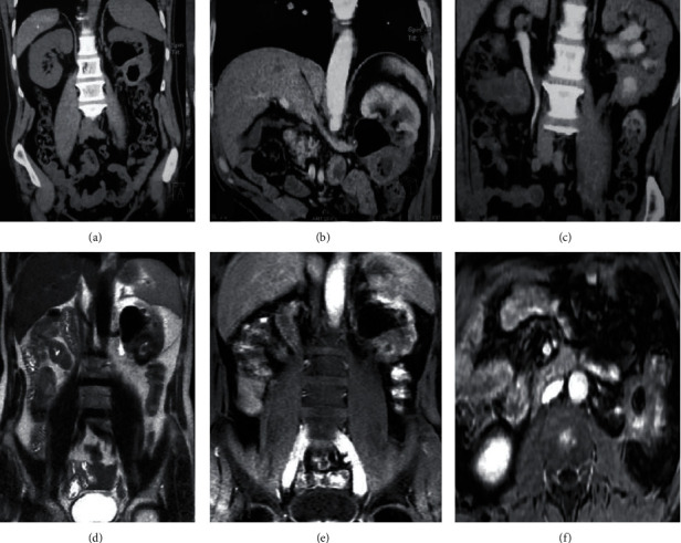
(a–c) Computed tomography; (a) pelvic hydroaeric level in the left kidney; (b) poorly enhanced lower pole in the left kidney; (c) contrast medium extending to the left colon. (d–f) Magnetic resonance imaging; (d) 41 × 38 × 41 mm T2 hypointense lower pole left renal mass associated with a pelvic hydroaeric level; (e) contrast medium extending to the left colon; (f) left renal mass invading the proximal ureter, the left colic angle, and the psoas muscle.
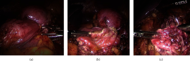
Laparoscopic images; (a) left renal tumor invading the left colon angle; (b) left lateral and segmental colonic resection began with opening the colonic mucosa in a healthy area; (c) closure of the colonic mucosa with a 4-0 vicryl thread.
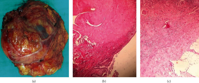
(a) Macroscopic image of the specimen; (b) inflammatory lesion infiltrating the intestinal mucosa (Hemalin-Eosine ×40); (c) renal tumor harboring an inflammatory lymphoplasmacytic infiltrate on a fibrous background (Hemalin-Eosine ×100).
References
-
- Coffin C. M., Watterson J., Priest J. R., Dehner L. P. Extrapulmonary inflammatory myofibroblastic tumor (inflammatory pseudotumor) a clinicopathologic and immunohistochemical study of 84 cases. The American Journal of Surgical Pathology . 1995;19(8):859–872. doi: 10.1097/00000478-199508000-00001. - DOI - PubMed
Publication types
LinkOut - more resources
Full Text Sources
