. 2022 Feb 21:12:8.
doi: 10.25259/JCIS_202_2021. eCollection 2022.

A case of spinal melorheostosis

Affiliations

A case of spinal melorheostosis

Alban Wei Shern Leong et al. J Clin Imaging Sci. .

Abstract

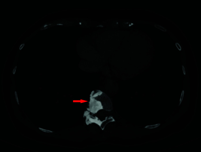
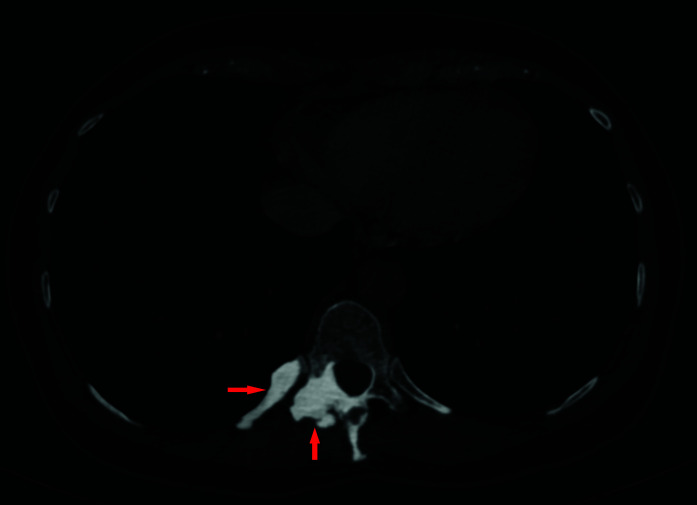
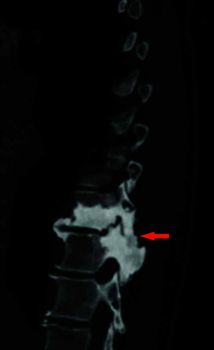
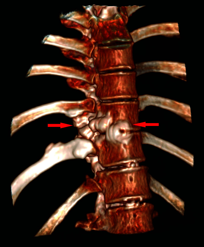
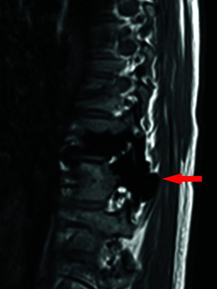
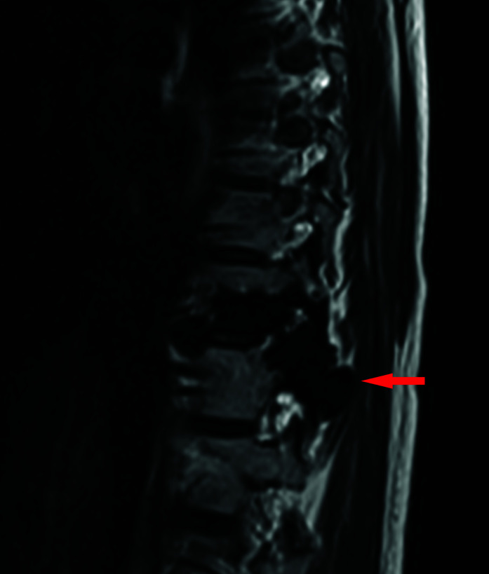
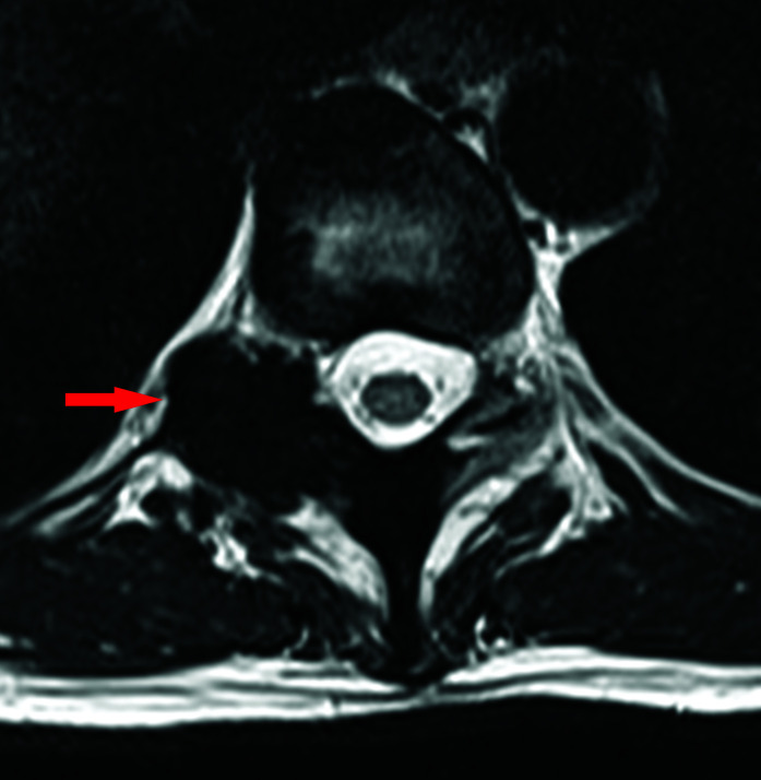
Melorheostosis is a rare bone dysplasia of unknown etiology with an incidence of 0.9 cases per million. It typically affects the upper or lower limbs and can cause severe deformity and functional impairment. Diagnosis is radiological and is often described as a "flowing candle wax" appearance on the radiograph. Treatment is individualized depending on the site and severity of symptoms. We report a rare case of spinal melorheostosis. We demonstrate the imaging features of melorheostosis on CT and MRI. We discuss the classification, genetics, and management of this condition.

Keywords: Asymptomatic; MAP2K1; Melorheostosis.


Conflict of interest statement

There are no conflicts of interest.

Figures

Figure 1A:
Figure 1A:
Axial CT image of the thorax in bone windows, showing a dense osteosclerotic lesion eccentrically located to the right of the T10 vertebral body with involvement of the right lamina. There is associated bone expansion with narrowing of the right neural exit foramen.
Figure 1B:
Figure 1B:
Axial CT image at a lower position compared to Figure 1A, also in bone windows, shows dense sclerotic changes also involving the adjacent T10 rib.
Figure 1C:
Figure 1C:
Sagittal CT image of the lower thorax, in bone windows, demonstrates contiguous osteosclerotic changes of the T10 and T11 vertebral bodies with mild bone expansion of the spinous process of T11.
Figure 1D:
Figure 1D:
Volume rendered CT image of the lower thoracic spine, shows the bony expansion of the vertebral bodies and ‘dripping candle wax’ appearance of the vertebra.
Figure 1E:
Figure 1E:
Sagittal T1 weighted images of the lower thoracic spine, demonstrating low signal change in the spine on both sequences, Figure 1E and 1F, consistent with a dense osteosclerotic bone lesion. There is involvement of the posterior elements and evidence of mild expansion.
Figure 1F:
Figure 1F:
Sagittal T2 weighted images of the lower thoracic spine, demonstrating low signal change in the spine on both sequences, Figure 1E and 1F, consistent with a dense osteosclerotic bone lesion. There is involvement of the posterior elements and evidence of mild expansion.
Figure 1G:
Figure 1G:
Axial T2 weighted image at the level of the T10-11 disc, shows loss of signal in the right posterior elements of T11 with bony expansion and narrowing of the right neural exit foramen.

References

    1. Leri A, Joanny J. Une afection non décrite des os hyperostose “en coulée” sur toute la longeur d’un member ou “melorhéostose”. Bull Mem Soc Med Hop Paris. 1922;46:1141–45. French.
    1. Clifford PD, Jose J. Melorheostosis. Am J Orthop (Belle Mead NJ) 2009;38:360–61. - PubMed
    1. Long HT, Li KH, Zhu Y. Severe Melorheostosis Involving the Ipsilateral Extremities. Clin Orthop Relat Res. 2009;467:2738–43. doi: 10.1007/s11999-009-0890-y. - DOI - PMC - PubMed
    1. Hellemans J, Preobrazhenska O, Willaert A, Debeer P, Verdonk PCM, Costa T et al. Loss-of-function mutations in LEMD3 result in osteopoikilosis, Buschke-Ollendorff syndrome and melorheostosis. Nat Genet. 2004;36:1213–18. doi: 10.1038/ng1453. - DOI - PubMed
    1. Kang H, Jha S, Deng Z, Fratzl-Zelman N, Cabral WA, Ivovic A et al. Somatic activating mutations in MAP2K1 cause melorheostosis. Nat Commun. 2018;9:1390. doi: 10.1038/s41467-018-03720-z. - DOI - PMC - PubMed

LinkOut - more resources