Case Reports
. 2022 Jan 30;14(1):e21727.
doi: 10.7759/cureus.21727. eCollection 2022 Jan.

Desmoid Tumors Arising on the Mesenteric Surgical Scar of Abdominal Sarcomas

Affiliations
Case Reports

Desmoid Tumors Arising on the Mesenteric Surgical Scar of Abdominal Sarcomas

Giovanni Damiani et al. Cureus. .

Abstract

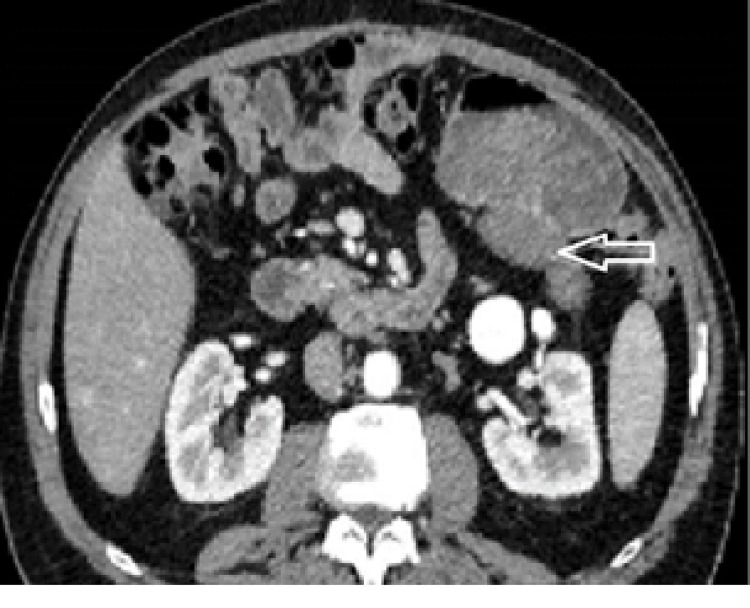
A sporadic desmoid tumor (DT) is a rare type of tumor of the mesenchymal connective tissues is now considered an intermediate disease or locally aggressive. They may develop on scars or after traumatism, favored by growth factors released during the initial phase of wound healing. Most of the abdominal DT arising on a scar is described on the wall incision. In this report, we describe two cases of DT arising on the intraperitoneal surgical scar, shortly after the resection of a low-grade retroperitoneal liposarcoma and a low-risk gastric gastrointestinal stromal tumor (GIST). Inconsistency between low risk according to the classification of the primary sarcoma and early local recurrence (LR) should raise the possibility of DT. Core needle biopsy (CNB) should be performed when it is feasible, including on local recurrences (LR). Surveillance has become the first-line treatment for DT. In case of progression between two imaging during the surveillance phase, surgery, when it's not mutilating, is indicated for selected cases as second-line treatment.

Keywords: desmoid tumor; mesenteric tumor; oncologic surgery; retroperitoneal tumor; sarcoma surgery; soft tissue tumor.


Conflict of interest statement

The authors have declared that no competing interests exist.

Figures

Figure 1
Figure 1. Case 1. Contrast-enhanced abdominal CT scan
This shows a retroperitoneal adipocytic mass displacing the right colon and kidney to the left (red arrow).
Figure 2
Figure 2. Case 1. Well-differentiated liposarcoma as the primary tumor
G stands for magnification (A) Gx10 tumor is composed of mature adipocytes with significant variation in cell size and of atypical hyperchromatic stromal cells admixed within a collagenous background. (B) Interphase fluorescence in situ hybridization using probes for MDM2 (red signal) and the centromere of chromosome 12 (green signal) showed high-level amplification of MDM2 grouped in clusters. MDM2 staining Gx10, the tumor cells showed a moderate nuclear MDM2 staining consistent with a well-differentiated liposarcoma. (C) The Desmoid tumor was made of bland myofibroblasts arranged in long fascicles with a fibrous stroma. (D) β-catenin-staining Gx20 showed a diffuse and strong cytoplasmic and nuclear overexpression, consistent with the diagnosis of DT.
Figure 3
Figure 3. Case 1. Contrast-enhanced abdominal CT scan
This shows a well-defined homogenous non-adipocytic round mass in the mesentery close to the staple line of the ileocolic anastomosis (red arrow).
Figure 4
Figure 4. Case 2. Contrast-enhanced abdominal CT scan
This shows a heterogeneous mass on the posterior wall of the antrum, part of the stomach (the white cross inside the tumor is to show the borders of the tumor because it is heterogeneous).
Figure 5
Figure 5. Case 2. GIST epithelioid type harboring a PDGFRA mutation
G: stands for magnification; GIST: gastrointestinal stromal tumor (A) Gx 0.46, well-circumscribed mass infiltrated the muscular wall (asterisk) and the subserosa of the stomach. (B) Gx5 tumor was made of spindle and epithelioid cells within a myxoid stroma. (C) CD117 staining, Gx20: PDGFRA_mutant GIST showed a characteristic limited and slight expression of KIT. (D) DOG1 staining, Gx20 diffuse expression of DOG1 confirmed the diagnosis of PDGFRA-mutant GIST. Desmoid tumor (E) Gx0, 35: low-power examination revealed a poorly circumscribed lesion with ill-defined infiltration of the subserosa and the muscular wall (asterisk) of the colon. (F) High-power examination revealed bland myofibroblasts without cytonuclear atypia and without necrosis, dispersed within a fibrous stroma. (G) β-catenin staining Gx20 by immunochemistry, tumor cells showed a diffuse and strong nuclear expression of β-catenin, consistent with the diagnosis of desmoid tumor. (H): C-KIT staining Gx10 by immunochemistry: conversely, to a specimen of the primary, tumor cells do not express anymore C-KIT. (I): DOG1 staining Gx10: Conversely to the specimen of the primary, tumor cells do not express any more DOG1.
Figure 6
Figure 6. Case 2. Contrast-enhanced abdominal CT scan
The white arrow points to a homogenous mass on the posterior wall of the stomach.

References

    1. World Health Organization. Lyon, France: IARC Press; [ Aug; 2021 ]. 2020. Soft Tissue and Bone Tumours. WHO Classification of Tumours, 5th Edition, Volume 3.
    1. The management of desmoid tumours: a joint global consensus-based guideline approach for adult and paediatric patients. Desmoid Tumor Working Group. Eur J Cancer. 2020;127:96–107. - PubMed
    1. Extra-abdominal primary fibromatosis: aggressive management could be avoided in a subgroup of patients. Bonvalot S, Eldweny H, Haddad V, et al. Eur J Surg Oncol. 2008;34:462–468. - PubMed
    1. Spontaneous regression of primary abdominal wall desmoid tumors: more common than previously thought. Bonvalot S, Ternès N, Fiore M, et al. Ann Surg Oncol. 2013;20:4096–4102. - PubMed
    1. Soft tissue and visceral sarcomas: ESMO-EURACAN-GENTURIS Clinical Practice Guidelines for diagnosis, treatment and follow-up. Gronchi A, Miah AB, Dei Tos AP, et al. Ann Oncol. 2021;32:1348–1365. - PubMed

Publication types

LinkOut - more resources