. 2022 Feb 25:2022:3686863.
doi: 10.1155/2022/3686863. eCollection 2022.

Immunohistochemical Analysis of Postburn Scars following Treatment Using Dermal Substitutes

Affiliations

Immunohistochemical Analysis of Postburn Scars following Treatment Using Dermal Substitutes

Mi Young Lee et al. Anal Cell Pathol (Amst). .

Abstract

Background: Post-burn hypertrophic scars commonly occur after burns. Studies that compare dermal substitutes with other treatment methods are insufficient. The purpose was to analyze the histopathological differences in hypertrophic burn scars after Matriderm®+split-thickness skin graft (STSG) and compare with AlloDerm®+STSG, STSG, full-thickness skin graft (FTSG), and normal skin.

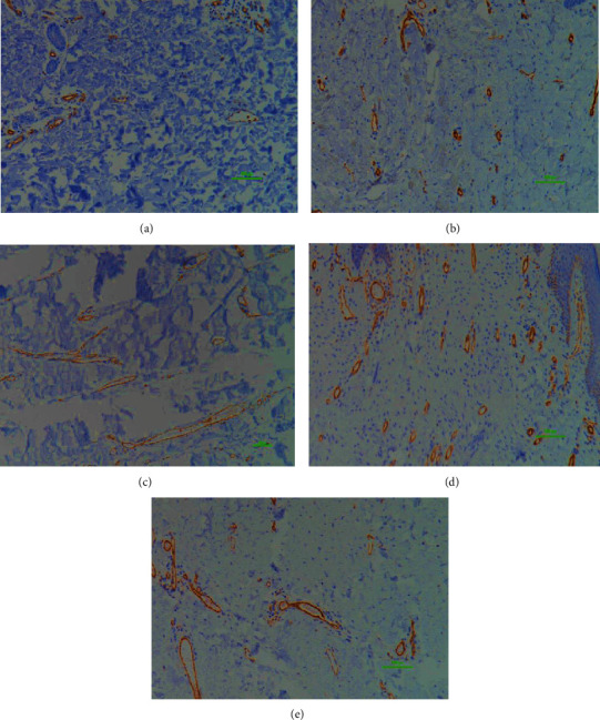
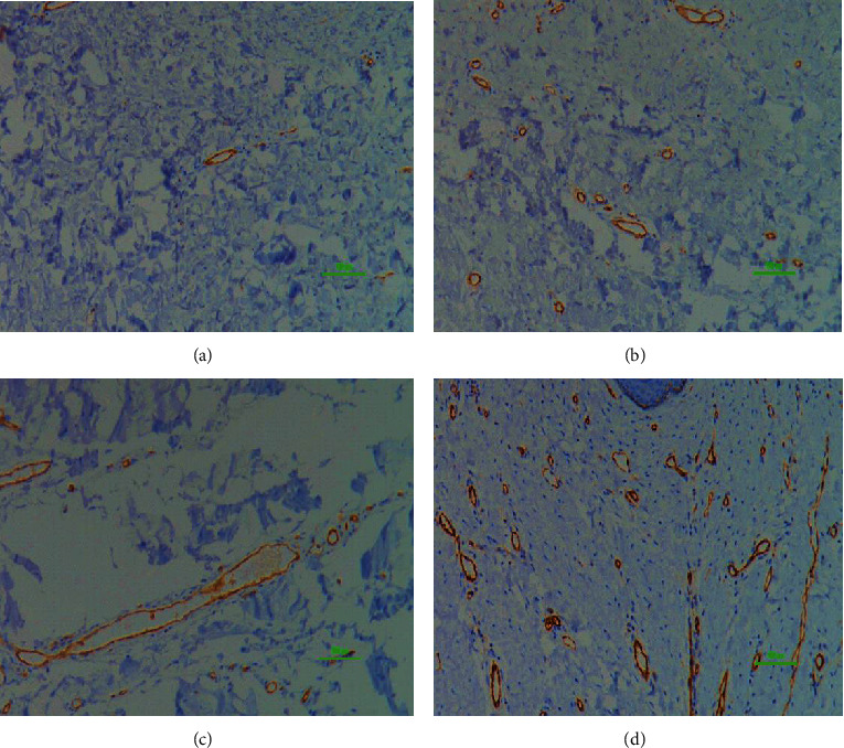
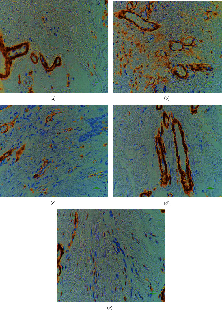
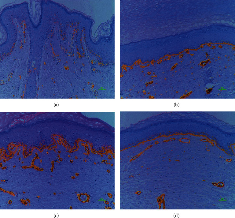
Methods: Samples of unburned, normal skin and deep 2nd or 3rd degree burns were obtained from patients who experienced a burn injury in the past to at least 6 months before biopsy, which was performed between 2011 and 2012. All subjects received >6 months of treatment before the biopsy. Intervention groups were normal (63), STSG (28), FTSG (6), Matriderm® (11), and AlloDerm® (18). Immunohistochemical analyses of elastin, collagen I, collagen III, cluster of differentiation 31 (CD31), smooth muscle actin (α-SMA), and laminin from scar and control tissues were performed and compared.

Results: α-SMA vascular quantity and vessel width, stromal CD31, and basement membrane laminin expression were not significantly different between normal and intervention groups. Matriderm® group showed no significant difference in elastin, collagen III, stromal CD31 and α-SMA, CD31 vessel width, stromal α-SMA, vessel quantity and width, and laminin length compared to the normal group, meaning they were not significantly different from the normal skin traits.

Conclusion: Dermal substitutes may be an optimal alternative to address the cosmetic and functional limitations posed by other treatment methods.


Conflict of interest statement

None declared.

Figures

Figure 1
Figure 1
Study flow diagram. Details of the study selection process.
Figure 2
Figure 2
Comparison of elastin fibers. Comparison of elastin fibers of the normal (a), STSG (b), and AlloDerm® (c) groups. 200x magnification, 100 μm scale bar; immunostaining technique. Elastic fibers are responsible for resilience and recoil in many tissues. There was a significant decrease in elastin fibers in the STSG and AlloDerm® groups compared to the normal group. Therefore, scar tissues in these two groups may be stiffer compared to the other groups.
Figure 3
Figure 3
Comparison of collagen III fibers. Collagen III fibers of the normal (a) and STSG (b) group; 200x magnification, 100 μm scale bar; immunostaining technique; Collagen provides tensile strength but hypertrophic scars contain an overabundance of collagen, contributing to a raised appearance and stiffness. There was a significant decrease in collagen III in the STSG group compared to the normal group.
Figure 4
Figure 4
Comparison of CD31 vessel quantity. CD31 vessel quantity in the normal (a), STSG (b), FTSG (c), Matriderm® (d), and AlloDerm® (e) groups, respectively; 100x magnification, 100 μm scale bar; immunostaining technique; Hyperactive fibroblasts in hypertrophic scars secrete higher than normal levels of angiogenic factors, which promote endothelial cell proliferation and more microvessel formation. CD31 helps to promote vascular barrier function in response to inflammatory stimuli. There was a significant increase in vessel quantity expressing CD31 in the FTSG and Matriderm® groups compared to the normal group.
Figure 5
Figure 5
Comparison of CD31 vessel length. CD 31 vessel length of the normal (a), STSG (b), FTSG (c), Matriderm® (d), and AlloDerm® (e) groups; 100x magnification, 100 μm scale bar; immunostaining technique; local collagen overproduction by hypertrophic scar fibroblasts mechanically squeezes the microvessels, thus leading to narrowing and deformation. CD31 helps to promote vascular barrier function in response to inflammatory stimuli. There was a significant increase in CD31 impregnated vessel length in the STSG, Matriderm®, and AlloDerm® groups compared to the normal group.
Figure 6
Figure 6
Comparison of CD31 vessel width. Comparison between control (a), STSG (b), FTSG (c), and Matriderm® (d); 100x magnification, 100 μm scale bar; immunostaining technique; local collagen overproduction by hypertrophic scar fibroblasts mechanically squeezes the microvessels, thus leading to narrowing and deformation. CD31 helps to promote vascular barrier function in response to inflammatory stimuli. There was a significant increase in CD31 impregnated vessel width in the FTSG compared to the control, STSG, and Matriderm® groups.
Figure 7
Figure 7
Comparison of α-SMA stroma. α-SMA of the normal (a), STSG (b), FTSG (c), Matriderm® (d), and AlloDerm® (e) group; 400x magnification, 100 μm scale bar; immunostaining technique; Myofibroblasts express α-SMA, and persistence of myofibroblasts may lead to excess scarring, which impairs function and aesthetics. There was a significant increase in α-SMA in the STSG, FTSG, and AlloDerm® compared with the normal and Matriderm® groups.
Figure 8
Figure 8
Comparison of laminin basement membrane intensity. Intensity of laminin in the basement membrane of the normal (a), STSG (b), Matriderm® (c), and AlloDerm® groups (d); 400x magnification, 100 μm scale bar; immunostaining technique; laminins have a central role in formation, the architecture, and the stability of basement membranes, thus making them essential for basement membrane assembly. There was a significant increase in laminin expression intensity in the STSG, Matriderm®, and AlloDerm® groups compared with the normal group.

Similar articles

Cited by

References

    1. Bombaro K. M., Evngrav L. H., Carrougher G. J., et al. What is the prevalence of hypertrophic scarring following burns? Burns . 2003;29(4):299–302. doi: 10.1016/S0305-4179(03)00067-6. - DOI - PubMed
    1. Finnerty C. C., Jeschke M. G., Branski L. K., Barret J. P., Dziewulski P., Herndon D. N. Hypertrophic scarring: the greatest unmet challenge after burn injury. Lancet . 2016;388(10052):1427–1436. doi: 10.1016/S0140-6736(16)31406-4. - DOI - PMC - PubMed
    1. Brissett A. E., Sherris D. A. Scar contractures, hypertrophic scars, and keloids. Facial Plastic Surgery . 2001;17(4):263–272. doi: 10.1055/s-2001-18827. - DOI - PubMed
    1. Clark J. A., Cheng J. C. Y., Leung K. S. Mechanical properties of normal skin and hypertrophic scars. Burns . 1996;22(6):443–446. doi: 10.1016/0305-4179(96)00038-1. - DOI - PubMed
    1. Deitch E. A., Wheelahan T. M., Rose M. P., Clothier J., Cotter J. Hypertrophic burn Scars. The Journal of Trauma . 1983;23(10):895–898. doi: 10.1097/00005373-198310000-00009. - DOI - PubMed