

Primary retroperitoneal extraovarian granulosa cell tumor
- PMID: 35252048
- PMCID: PMC8893158
- DOI: 10.4322/acr.2021.355
Primary retroperitoneal extraovarian granulosa cell tumor
Abstract
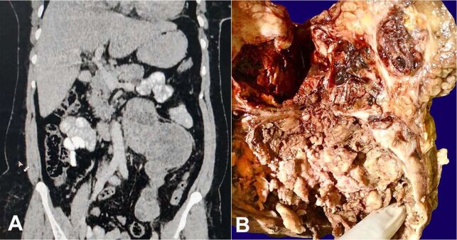
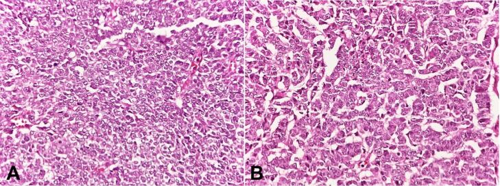
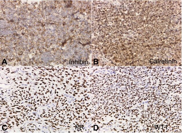
Extraovarian granulosa cell tumors (GCTs) develop from ectopic gonadal tissue situated along the embryonal route of the genital ridge. Primary retroperitoneal tumors are extremely rare, with an incidence of 02% -06% and 80-85% probability of malignancy. Only eight such case reports have been published previously. We herein, report a rare case of extraovarian retroperitoneal GCT in a 55-year-old woman who presented with intermittent left lumbar region pain of one-year duration. She had a history of hysterectomy and bilateral salpingo-oophorectomy 8 years ago for uterine leiomyoma. Laparotomy revealed a retroperitoneal mass measuring 8cm x 10cm x 20cm in size, solid cystic with areas of necrosis and hemorrhage. The gross features, classical histopathology, and positive immunostaining of the retroperitoneal mass with inhibin, calretinin, PR, WT1 and immunonegativity for EMA were characteristic of adult-type GCT. Excluding any previous history of primary ovarian GCT in this patient, a de-novo retroperitoneal diagnosis was established.
Keywords: Granulosa cell tumor; immunohistochemistry; inhibins.
Copyright © 2022 The Author(s).
Conflict of interest statement
Conflict of interest: None.
Figures
References
-
- Vassallo MC, Collict M, Buhagiar T, Formosa M. Retroperitoneal granulosa cell tumor. J Case Rep Images Obstet Gynecol. 2019;5:100045Z08MV2019
-
- Swain SK, Ahmed A, Mohan AT, Munikrishnan V. Primary extraovarian Granulosa Cell Tumor (GCT) of Omentum: a rare occurrence. Trop Gastroenterol. 2020;41(2):48–50.
Publication types
LinkOut - more resources
Full Text Sources
Research Materials
