. 2022 Feb 21;8(3):e08988.
doi: 10.1016/j.heliyon.2022.e08988. eCollection 2022 Mar.

Labyrinthin: A distinct pan-adenocarcinoma diagnostic and immunotherapeutic tumor specific antigen

Affiliations

Labyrinthin: A distinct pan-adenocarcinoma diagnostic and immunotherapeutic tumor specific antigen

Michael Babich et al. Heliyon. .

Abstract

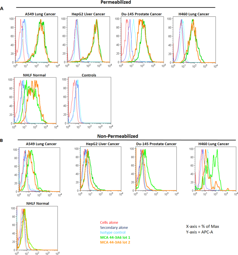
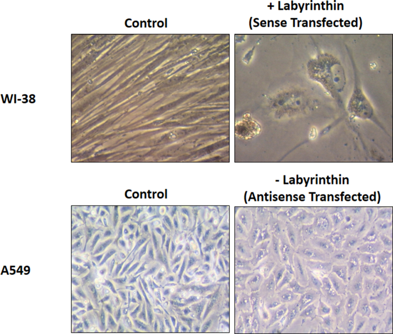
Structural analysis and detection of optimal cell surface localization of labyrinthin, a pan-adenocarcinoma target, was studied with respect to adenocarcinoma specificity vs. normal and non-adenocarcinoma cells. Patient-derived tissue microarray immunohistochemistry (IHC) was performed on 729 commercially prepared tissue blocks of lung, colon, breast, pancreas, prostate, and ovary cancers combined, plus a National Cancer Institute (NCI) tissue microarray derived from another 236 cases. The results confirmed that anti-labyrinthin mouse monoclonal MCA 44-3A6 antibody recognized adenocarcinomas, but not normal or non-adenocarcinoma cancer cells. The consensus of multiple topology analysis programs on labyrinthin (255 amino acids) estimate a type II cell membrane associated protein with an N-terminus signal peptide. However, because the labyrinthin sequence is enveloped within the 758 amino acids of the intracellular aspartyl/asparaginyl beta-hydroxylase (ASPH), a purported tumor associated antigen, standard IHC methods that permeabilize cells can expose common epitopes. To circumvent antibody cross-reactivity, cell surface labyrinthin was distinguished from intracellular ASPH by FACS analysis of permeabilized vs non-permeabilized cells. All permeabilized normal, adeno-and non-adenocarcinoma cells produced a strong MCA 44-3A6 binding signal, likely reflecting co-recognition of intracellular ASPH proteins along with internalized labyrinthin, but in non-permeabilized cells only adenocarcinoma cells were positive for labyrinthin. Confocal microscopy confirmed the FACS results. Labyrinthin as a functional cell-surface marker was suggested when: 1) WI-38 normal lung fibroblasts transfected with labyrinthin sense cDNA displayed a cancerous phenotype; 2) antisense transfection of A549 human lung adenocarcinoma cells appeared more normal; and 3) MCA44-3A6 suppressed A549 cell proliferation. Collectively, the data indicate that labyrinthin is a unique, promising adenocarcinoma tumor-specific antigen and therapeutic target. The study also raises a controversial issue on the extent, specificity, and usefulness of ASPH as an adenocarcinoma tumor-associated antigen.

Keywords: ASPH; Adenocarcinoma; Junctate; Labyrinthin; Neoantigen; Pan-tumor target; Tumor associated antigen; Tumor specific antigen.


Conflict of interest statement

Michael Babich and James Radosevich are principals, and Ankit Sharma is a research fellow, at LabyRx Immunological Therapeutics (USA) Limited.

Figures

Figure 1
Figure 1
Detection of labyrinthin on human tissue array by immunohistochemistry. Labyrinthin expression (brown stain) as detected by MCA 44-3A6 on cancer tissue arrays. Pictures are representative from normal or malignant samples [ ( ) = number of samples in the block]. Omission of the primary antibody or substitution of a non-relevant primary antibody did not produce specific immunostaining reactions (data not shown). ∗Nomenclature: Normal = FDA991 block; Malignant = LC1002, lung cancer; CO208, colon cancer; etc. for the respective corresponding blocks in each panel. Magnification 20x.
Figure 2
Figure 2
Computer-based determination of labyrinthin and ASPH cellular localization. Depiction of labyrinthin (left) localized on the extracellular side as predicted by the Protter program. A TM domain has been reported by some programs [43]; Protter found TM that was indeterminant but did possess a signal peptide (amino acids 1–17; coded in red). By comparison, no signal peptide was found for ASPH, but a transmembrane domain (amino acids 54–74) was identified for intracellular organelle-associated ASPH (right).
Figure 3
Figure 3
FACS analysis of MCA 44-3A6 epitopes in permeabilized vs. intact cells. Normal (NHLF) human lung fibroblasts and adenocarcinoma (all other) cells were immunolabeled with two different MCA 44-3A6 antibody preparations (Lot 1: commercially grown; Lot 2: grown in the present lab). (A) Permeabilized cells all displayed a significant shift to either anti-labyrinthin antibody preparation. (B) Rightward shift displayed by intact adenocarcinoma cells (A549, H460, HepG2); Du-145 cells displayed less of a response to Lot 1 antibody. Results are representative of at least 2 different preparations. Isotype control: mouse IgG; secondary alone: Alexa647 anti-mouse IgG.
Figure 4
Figure 4
Cell surface localization of labyrinthin by single cell image analysis. Immunolabeled MCA 44-3A6 antibody (Alexa 488; green) binding to A549 and WI-38 cells was detected by confocal microscopy. The images shown include differential interference contrast (DIC) to visualize unstained, transparent samples; DAPI and Alexa 488 to visualize nuclear and labyrinthin localization; and overlay of the two preceding images. (A) Permeabilized A549 and WI-38 cells. All cells for each of the preparations displayed a positive signal. (B) Representative images of intact, non-permeabilized cells in which all A549 cells had punctate antibody binding on their surfaces, whereas no signal was detected in WI-38 cells. Data are representative from at least two preparations; magnification 40x.
Figure 5
Figure 5
Effects of labyrinthin over- or under-expression on cell morphology. Phase contrast microscopy (x20) of normal WI-38 human lung fibroblasts and A549 human lung adenocarcinoma cells transfected with the full length labyrinthin sense- or antisense-cDNA constructs, respectively. WI-38 control cells were transfected with pBK-CMV plasmid alone. No effect of mock transfected was detected compared to untreated WI-38 control cells (not shown). A549 control and minus (-) labyrinthin cells were transfected with pBK-CMV plasmid sense- and antisense-cDNA, respectively. No effect was observed between control labyrinthin overexpression (as shown) vs. untreated A549 cells (not shown).
Figure 6
Figure 6
Anti-labyrinthin antibody inhibition of A549 cell proliferation. Ascitic fluid containing the given amounts of protein from control (SP2/0 cell line) or MCA 44-3A6 hybridomas was added to 80–90% confluent cells. Three days later cell proliferation was measured by MTT assay. Lower amounts of ascites (≤3.125 ug/mL) were without effect compared to no addition (not shown). N ≥ 8 per treatment; significant difference between control vs. treatment lines (p ≤ 0.01).

References

    1. Babich M., Sharma A., Lee T., Radosevich A.J. Topology and adenocarcinoma cell localization dataset indicate labyrinthin as a neoantigen. Data Brief. 2021 submitted for publication.
    1. Urbanek-Trzeciak M.O., Galka-Marciniak P., Nawrocka P.M., Kowal E., Szwec S., Giefing M., Kozlowski P. Pan-cancer analysis of somatic mutations in miRNA genes. EBioMedicine. 2020;61:103051. - PMC - PubMed
    1. Ma W., Pham B., Li T. Cancer neoantigens as potential targets for immunotherapy. Clin. Exp. Metastasis. 2021:1–10. - PMC - PubMed
    1. Looney A.M., Nawaz K., Webster R.M. Tumour-agnostic therapies. Nat. Rev. Drug Discov. 2020;19(6):383–385. - PubMed
    1. Li L., Goedegebuure S.P., Gillanders W.E. Preclinical and clinical development of neoantigen vaccines. Ann. Oncol. 2017;28:xii11–xii17. - PMC - PubMed