Comment
. 2022 Feb 26:17:26331055221080175.
doi: 10.1177/26331055221080175. eCollection 2022.

Brain Networks of Connectionally Unique Basolateral Amygdala Cell Types

Affiliations
Comment

Brain Networks of Connectionally Unique Basolateral Amygdala Cell Types

Houri Hintiryan et al. Neurosci Insights. .

Abstract

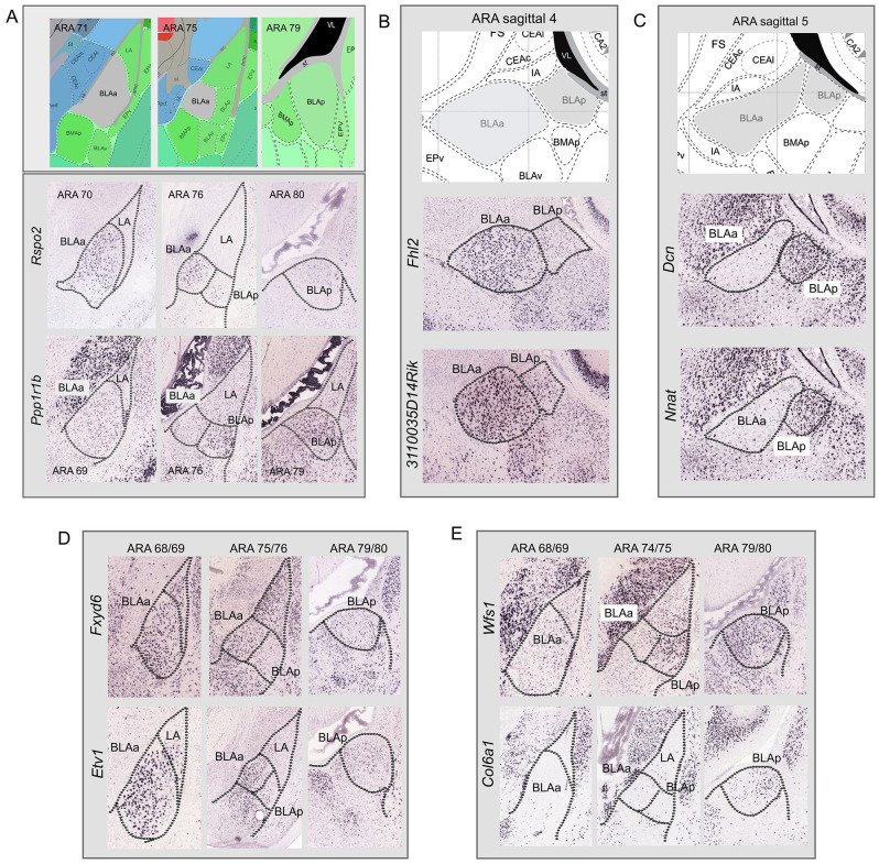
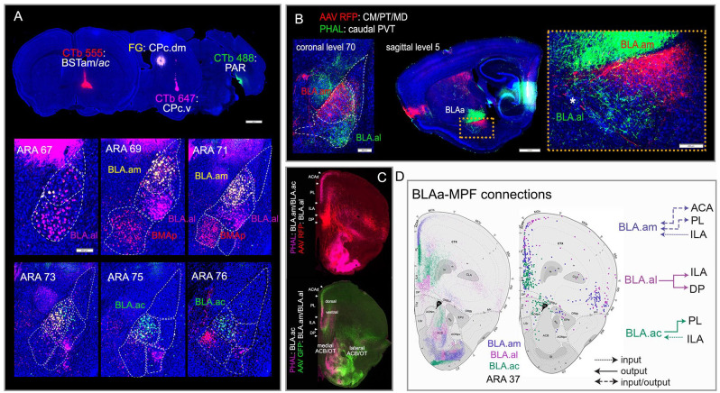
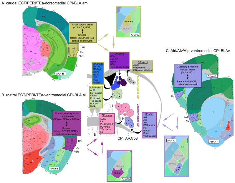
Different brain regions structurally interconnected through networks regulate behavior output. Therefore, understanding the functional organization of the brain in health and disease necessitates a foundational anatomic roadmap to its network organization. To provide this to the research community, our lab has systematically traced thousands of pathways in the mouse brain and has applied computational measures to determine the network architecture of major brain systems. Toward this effort, the brain-wide networks of the basolateral amygdalar complex (BLA) were recently generated. The data revealed uniquely connected cell types within the same BLA nucleus that were constituents of distinct neural networks. Here, we elaborate on how these connectionally unique BLA cell types fit within the larger cortico-basal ganglia and limbic networks that were previously described by our team. The significance and utility of high quality, detailed anatomic data is also discussed.

Keywords: Basolateral amygdala; brain networks; cell types; circuit tracing; connectome; neuroanatomy.


Conflict of interest statement

Declaration of Conflicting Interests: The author(s) declared no potential conflicts of interest with respect to the research, authorship, and/or publication of this article.

Figures

Figure 1.
Figure 1.
Gene expression in BLAa and BLAp. (A) In situ hybridization images from the Allen Brain Atlas, a genome wide gene expression atlas, showing that magnocellular BLAa neurons express Rspo2, while parvocellular BLAp neurons express primarily Ppp1r1b. Additional genes highlight the genetic similarity within the BLAa and their distinction from BLAp. (B and D) Genes Fhl2, 3110035D14Rik, Fxyd6, and Etv1, among others not shown here, are expressed primarily in BLAa and not BLAp. (C and E) Genes Dcn, Nnat, Wfs1, and Col6a1, among others not shown here, are expressed primarily in BLAp and not BLAa.
Figure 2.
Figure 2.
Connectionally unique BLAa cell types and their connectivity with medial prefrontal cortex (MPF). (A) Four retrograde tracer injections placed in BSTam (CTb 555: red), dorsomedial caudal CP (FG: yellow), ventrolateral caudal CP (CTb 647: pink), and in PAR (CTb 488: green) clearly reveal uniquely connected projection neurons in the three BLAa domains: BLA.am (FG), BLA.al (CTb 647), and BLA.ac (CTb 488). BMAp neurons that target BST are also labeled (CTb 555). Note the segregation between FG (yellow) and CTb 647 (pink) labeled cells in BLA.am and BLA.al, respectively at ARA levels 69 and 71. Also note the absence of CTb 488 (green) labeled cells in BLAa at ARA levels 67-71. (B) Anterograde tracers AAV-RFP and PHAL injected in different thalamic nuclei distinctly label BLA.am and BLA.al, which is evident in coronal (left) and sagittal (right) planes. Inset shows magnified version of boxed BLAa region on sagittal section. *Denotes boundary between BLAa and IA (intercalated amygdalar nucleus). (C) Top panel: Anterograde labeling from tracer injections made primarily in BLA.am (PHAL: pink) and BLA.al (AAV RFP: red) shows their topographic projections to MPF. Bottom panel: Anterograde labeling from tracer injections made primarily in BLA.ac (PHAL: pink) and BLA.al (AAV GFP: green) shows their distinct connections with MPF, ACB, OT, and CP. Note the stronger projections from BLA.am to dorsal PL/ACA compared to projections from BLA.ac to more ventral parts of PL, which is also evident in D (left; purple and green fibers in PL). (D) Schematic summarizing how each BLAa domain shares unique input/output connections with MPF, especially layers 2/3. Left shows projections from the BLAa domains to MPF, while the right shows MPF cells that project to BLAa domains. Note (1) the reciprocal connections between the BLA.am and ACA and PL (layer 2), (2) that the BLA.al mostly projects to ILA, and (3) the unique BLA.ac connection in which it heavily projects to PL (layer 2) but receives strong input from ILA. Source: Figure adapted from Hintiryan et al. Abbreviations: ac, anterior commissure; ACA, anterior cingulate cortex; ACB, nucleus accumbens; BSTam, anteromedial bed nucleus of stria terminalis; CM, central medial thalamic nucleus; CP, caudoputamen; ILA, infralimbic cortex; MD, mediodorsal thalamic nucleus; OT, olfactory tubercle; PAR, parasubiculum; PL, prelimbic cortex; PT, parataenial thalamic nucleus; PVT, paraventricular thalamic nucleus.
Figure 3.
Figure 3.
Visual representations of cortico-striatal-BLA subnetworks. The intermediate CP (CPi) consists of 4 major divisions, each of which receives and integrates different cortical input. These are (1) the dorsomedial, (2) ventromedial, (3) dorsolateral, and (4) ventrolateral. Note that only the dorsomedial (yellow) and ventromedial (magenta) divisions are shown. Based on highly topographic cortico-striatal projections, each division can be subdivided into domains, smaller regions that receive convergent input from different cortical areas. Adjacent domains, particularly within a community, share integrated cortical information (for details, see Hintiryan et al). Each circle on the CPi denotes the center of terminations from a single cortical area. A group of color-coded circles represents a domain. Color-coded boxes list cortical areas that project to that domain (name of domain included, e.g., CPi.dm.dm). The stroke color of the boxes denotes the larger CPi division within which the domains interact (yellow: dorsomedial CPi; magenta: ventromedial CPi). (A) Visual representation of the caudal ECT/PERI/TEa-dorsomedial CPi-BLA.am subnetwork showing the interactions of these 3 regions potentially involved in visual information processing. At the cortical level, the caudal ECT/PERI/TEa is heavily interconnected with visual processing areas. The caudal ECT/PERI/TEa project to the dorsomedial CPi (stroke colored yellow), which also receives input from a wide variety of visual processing cortical areas. It also is the region of the CPi that BLA.am cells target. The caudal ECT/PERI/TEa also provides input to BLA.am. (B) Visual representations of the rostral ECT/PERI/TEa-ventromedial CPi-BLA.al subnetwork showing interactions among these three regions potentially involved in gustatory/visceral information processing. At the cortical level, the rostral ECT/PERI/TEa is heavily interconnected with somatic sensory motor regions that regulate the mouth. Rostral ECT/PERI/TEa project to the ventromedial CPi (stroke colored magenta), where cortical gustatory and visceral information is integrated. This is also the region in the CPi that BLA.al cells target. The rostral ECT/PERI/TEa also provides input to the BLA.al. (C) Visual representation of the AId/AIv/AIp-ventromedial CPi-BLAv subnetwork. The AId/AIv/AIp receives input from gustatory and visceral cortical areas and projects to the ventromedial CPi, which also receives input from cortical regions that process the same type of information. The BLAv also projects to the same CPi region and the AId/AIv/AIp project to the BLAv. Abbreviations: ACA, anterior cingulate cortex; AId, agranular insular cortex, dorsal part; AIp, agranular insular cortex, posterior part; AIv, agranular insular cortex, ventral part; AUD, auditory cortex; CPi, intermediate caudoputamen; d, dorsal; ECT, ectorhinal cortex; ENT, entorhinal cortex; GU, gustatory cortex; i, intermediate; l, lateral; m, medial; ORB, orbitofrontal cortex; PERI, perirhinal cortex; PIR, piriform cortex; PTLp, posterior parietal association areas; RSP, retrosplenial cortex; TEa, posterior temporal association cortex; v, ventral; VIS, visual cortex; VISC, visceral cortex.

Comment on

  • Connectivity characterization of the mouse basolateral amygdalar complex.
    Hintiryan H, Bowman I, Johnson DL, Korobkova L, Zhu M, Khanjani N, Gou L, Gao L, Yamashita S, Bienkowski MS, Garcia L, Foster NN, Benavidez NL, Song MY, Lo D, Cotter KR, Becerra M, Aquino S, Cao C, Cabeen RP, Stanis J, Fayzullina M, Ustrell SA, Boesen T, Tugangui AJ, Zhang ZG, Peng B, Fanselow MS, Golshani P, Hahn JD, Wickersham IR, Ascoli GA, Zhang LI, Dong HW. Hintiryan H, et al. Nat Commun. 2021 May 17;12(1):2859. doi: 10.1038/s41467-021-22915-5. Nat Commun. 2021. PMID: 34001873 Free PMC article.

References

    1. Muñoz-Castañeda R, Zingg B, Matho KS, et al.. Cellular anatomy of the mouse primary motor cortex. Nature. 2021;598:159-166. - PMC - PubMed
    1. BRAIN Initiative Cell Census Network. A multimodal cell census and atlas of the mammalian primary motor cortex. Nature. 2021;598:86-102. - PMC - PubMed
    1. Peng H, Xie P, Liu L, et al.. Morphological diversity of single neurons in molecularly defined cell types. Nature. 2021;598:174-181. - PMC - PubMed
    1. Zhang Z, Zhou J, Tan P, et al.. Epigenomic diversity of cortical projection neurons in the mouse brain. Nature. 2021;598:167-173. doi:10.1038/s41586-021-03223-w - DOI - PMC - PubMed
    1. Hintiryan H, Bowman I, Johnson DL, et al.. Connectivity characterization of the mouse basolateral amygdalar complex. Nat Commun. 2021;12:2859. - PMC - PubMed

LinkOut - more resources