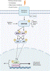
c-JUN n-Terminal Kinase (JNK) Signaling in Autosomal Dominant Polycystic Kidney Disease
- PMID: 35253003
- PMCID: PMC8896658
- DOI: 10.33696/Signaling.3.068
c-JUN n-Terminal Kinase (JNK) Signaling in Autosomal Dominant Polycystic Kidney Disease
Abstract
Polycystic kidney disease is an inherited degenerative disease in which the uriniferous tubules are replaced by expanding fluid-filled cysts that ultimately destroy organ function. Autosomal dominant polycystic kidney disease (ADPKD) is the most common form, afflicting approximately 1 in 1,000 people and is caused by mutations in the transmembrane proteins polycystin-1 (Pkd1) and polycystin-2 (Pkd2). The mechanisms by which polycystin mutations induce cyst formation are not well understood, however pro-proliferative signaling must be involved for tubule epithelial cell number to increase over time. We recently found that the stress-activated mitogen-activated protein kinase (MAPK) pathway c-Jun N-terminal kinase (JNK) pathway is activated in cystic disease and genetically removing JNK reduces cyst growth driven by a loss of Pkd2. This review covers the current state of knowledge of signaling in ADPKD with an emphasis on the JNK pathway.
Keywords: Cilia; Jun N Terminal kinase; Mitogen-activated protein kinase signaling; Mus musculus; Polycystic kidney disease; Polycystin-1; Polycystin-2.
Conflict of interest statement
Disclosures The authors have no conflicts to disclose.
Figures


Similar articles
-
c-Jun N-terminal kinase (JNK) signaling contributes to cystic burden in polycystic kidney disease.PLoS Genet. 2021 Dec 28;17(12):e1009711. doi: 10.1371/journal.pgen.1009711. eCollection 2021 Dec. PLoS Genet. 2021. PMID: 34962918 Free PMC article.
-
The genetics and physiology of polycystic kidney disease.Semin Nephrol. 2001 Mar;21(2):107-23. doi: 10.1053/snep.2001.20929. Semin Nephrol. 2001. PMID: 11245774 Review.
-
Mutant polycystin-2 induces proliferation in primary rat tubular epithelial cells in a STAT-1/p21-independent fashion accompanied instead by alterations in expression of p57KIP2 and Cdk2.BMC Nephrol. 2008 Aug 25;9:10. doi: 10.1186/1471-2369-9-10. BMC Nephrol. 2008. PMID: 18721488 Free PMC article.
-
Cell-Autonomous Hedgehog Signaling Is Not Required for Cyst Formation in Autosomal Dominant Polycystic Kidney Disease.J Am Soc Nephrol. 2019 Nov;30(11):2103-2111. doi: 10.1681/ASN.2018121274. Epub 2019 Aug 26. J Am Soc Nephrol. 2019. PMID: 31451534 Free PMC article.
-
Cilia and polycystic kidney disease.Semin Cell Dev Biol. 2021 Feb;110:139-148. doi: 10.1016/j.semcdb.2020.05.003. Epub 2020 May 28. Semin Cell Dev Biol. 2021. PMID: 32475690 Review.
Cited by
-
Illumination of understudied ciliary kinases.Front Mol Biosci. 2024 Mar 8;11:1352781. doi: 10.3389/fmolb.2024.1352781. eCollection 2024. Front Mol Biosci. 2024. PMID: 38523660 Free PMC article. Review.
-
Cilia Structure and Function in Human Disease.Curr Opin Endocr Metab Res. 2024 Mar;34:100509. doi: 10.1016/j.coemr.2024.100509. Epub 2024 Feb 20. Curr Opin Endocr Metab Res. 2024. PMID: 38836197 Free PMC article.
-
The Role of the Dysregulated JNK Signaling Pathway in the Pathogenesis of Human Diseases and Its Potential Therapeutic Strategies: A Comprehensive Review.Biomolecules. 2024 Feb 19;14(2):243. doi: 10.3390/biom14020243. Biomolecules. 2024. PMID: 38397480 Free PMC article. Review.
-
TNF-alpha promotes cilia elongation via mixed lineage kinases signaling in mouse fibroblasts and human RPE-1 cells.Cytoskeleton (Hoboken). 2024 Nov;81(11):639-647. doi: 10.1002/cm.21873. Epub 2024 May 20. Cytoskeleton (Hoboken). 2024. PMID: 38767050
-
Transforming advancing autosomal dominant polycystic kidney disease care: investigating new horizons in treatment and research.Inflammopharmacology. 2025 Aug 14. doi: 10.1007/s10787-025-01894-9. Online ahead of print. Inflammopharmacology. 2025. PMID: 40813527 Review.
References
Grants and funding
LinkOut - more resources
Full Text Sources
Research Materials
Miscellaneous
