. 2022 Apr;70(4):299-310.
doi: 10.1369/00221554221085336. Epub 2022 Mar 6.

Vascular E-selectin Expression Detected in Formalin-fixed, Paraffin-embedded Sections With an E-selectin Monoclonal Antibody Correlates With Ulcerative Colitis Activity

Affiliations

Vascular E-selectin Expression Detected in Formalin-fixed, Paraffin-embedded Sections With an E-selectin Monoclonal Antibody Correlates With Ulcerative Colitis Activity

Masataka Murahashi et al. J Histochem Cytochem. 2022 Apr.

Abstract

It is widely accepted that E-selectin, an inducible endothelial cell adhesion molecule, plays a critical role in the initial step of neutrophil recruitment to sites of acute inflammation. However, immunohistological analysis of E-selectin has been hampered by lack of E-selectin-specific monoclonal antibodies that can stain formalin-fixed, paraffin-embedded (FFPE) tissue sections. Here, we employed E-selectin•IgM (a soluble form of E-selectin) as immunogen, and then, after negative selection with L-selectin•IgM and P-selectin•IgM and screening of FFPE sections of both COS-1 cells overexpressing E-selectin and acute appendicitis tissues, we successfully generated an E-selectin-specific monoclonal antibody capable of staining FFPE tissue sections. We used this antibody, designated U12-12, to perform quantitative immunohistological analysis of 390 colonic mucosal biopsy specimens representing ulcerative colitis. We found that the higher the histological disease activity, the greater the number of vessels expressing E-selectin, an observation consistent with previous analyses of frozen tissue sections. Furthermore, in active ulcerative colitis, E-selectin-expressing vessels contained neutrophils attached to endothelial cells, presumably in the process of extravasation, which eventually could cause epithelial damage. These results overall indicate that U12-12 is effective for E-selectin immunohistochemistry in archived FFPE samples representing various human diseases.

Keywords: carbohydrate-binding protein; colon; inflammatory bowel disease; lectin; sialyl Lewis x.


Conflict of interest statement

Competing Interests: The author(s) declared no potential conflicts of interest with respect to the research, authorship, and/or publication of this article.

Figures

Figure 1.
Figure 1.
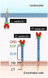
Schematic representation of the structure of E-selectin, L-selectin, and P-selectin. Each consists of five domains: (1) an N-terminal C-type lectin domain (also called the carbohydrate recognition domain [CRD]), (2) an epidermal growth factor (EGF)-like domain, (3) a set number of sequential short consensus repeats (SCRs, also called Sushi) domains, (4) a transmembrane (TM) domain, and (5) a C-terminal short cytoplasmic tail (CT). Note that both E-selectin and P-selectin are expressed on endothelial cells.
Figure 2.
Figure 2.
Immunocytological detection of E-selectin expressed in Chinese hamster ovary (CHO) cells. (A) Flow cytometry analysis of CHO/E-selectin cells performed with 68-5H11 (upper filled histogram) and U12-12 (lower filled histogram) antibodies. Unfilled histograms represent negative controls established in the absence of primary antibody. X- and y-axes indicate fluorescence intensity and number of events, respectively. Note that cells expressing E-selectin are distributed normally, indicating that they are of a single clone. (B) Immunofluorescence of formalin-fixed CHO/E-selectin cells stained with the U12-12 antibody. Nuclei were stained with DAPI. Bar = 50 µm. Abbreviation: DAPI, 4′,6-diamidino-2-phenylindole.
Figure 3.
Figure 3.
E-selectin-specific U12-12 binding. (A) Western blot analysis of lysates of COS-1 cells overexpressing human E-selectin using U12-12 (right lane). Lysates of mock-transfected COS-1 cells served as controls (left lane). (B) Western blot analysis of conditioned media of COS-1 cells transfected with cDNA encoding E-selectin•IgM, L-selectin•IgM, or P-selectin•IgM. Membranes were immunoblotted (IB’d) with anti-human IgM (left panel) or U12-12 (right panel).
Figure 4.
Figure 4.
Epitope mapping by domain deletion. (A) Schematic representation of an E-selectin•IgM chimera (wild-type) and corresponding domain deletion mutants (ΔCRD, ΔEGF, ΔSushi 1, and ΔSushi 2). A recombinant E-selectin Sushi 2 domain fused to IgM (Sushi 2), which does not contain the CRD, the EGF-like domain, or the Sushi 1 domain, was also constructed. Each mutant includes the wild-type signal peptide (SP) (dotted square on far left). The relative length of each domain has been adjusted to match the wild-type form, but the Fc region of IgM has been truncated. (B) Western blot analysis of deletion mutants shown in (A) immunoblotted (IB’d) with anti-human IgM (upper panels) or U12-12 (lower panels). (C) Double immunofluorescence staining of COS-1 cells expressing P-selectin•FLAG with anti-FLAG (left panel; red) or U12-12 (middle panel; secondary antibody Alexa Fluor 488 is not detectable). Merged images are shown at right. Nuclei were stained with DAPI. Bar = 50 µm. Abbreviation: CRD, carbohydrate recognition domain; EGF, epidermal growth factor; DAPI, 4′,6-diamidino-2-phenylindole.
Figure 5.
Figure 5.
Vascular expression of E-selectin in acute appendicitis. FFPE (upper panels) and frozen (lower panels) tissue sections of acute appendicitis were stained with hematoxylin and eosin (HE; left panels) or immunostained with U12-12 (middle and right panels). Images at right (upper and lower rows) are respective enlarged views of the region adjacent to the asterisk in the corresponding middle panel, viewed using an oil immersion lens. Signals were visualized with DAB (brown) and tissues were counterstained with hematoxylin. In right panels, note that neutrophils in the vascular lumen appear to attach to U12-12-positive endothelial cells (arrows). Bar = 50 µm for the left and middle panels and 10 µm for the right panel. Abbreviation: FFPE, formalin-fixed, paraffin-embedded.
Figure 6.
Figure 6.
Induction of E-selectin on venular endothelial cells in active ulcerative colitis. FFPE tissue sections of ulcerative colitis in either active phase (Geboes grade 5; upper panels) or remission phase (Geboes grade 1; lower panels) were stained with hematoxylin and eosin (HE; left panels) or immunostained with U12-12 (middle and right panels). Images at right (upper and lower rows) are respective enlarged views of the region adjacent to the asterisk in the corresponding middle panel, viewed using an oil immersion lens. Signals were visualized with DAB (brown) and tissues were counterstained with hematoxylin. In the right upper panel, note that neutrophils in the vascular lumen seen in active ulcerative colitis (Geboes grade 5) appear to attach to U12-12-positive endothelial cells (arrows). Bar = 50 µm for left and middle panels and 10 µm for right panels. Abbreviation: FFPE, formalin-fixed, paraffin-embedded.
Figure 7.
Figure 7.
Scatter plot representing one-way ANOVA of the number of U12-12-positive vessels (y-axis) based on Geboes grade (x-axis). In the case of multiple identical values, data points are superimposed, and outliers are omitted. The vertical span of each green diamond represents the 95% confidence interval (CI) for each group. The center line across each green diamond corresponds to the group mean. Abbreviation: ANOVA, analysis of variance.

References

    1. Wellicome SM, Thornhill MH, Pitzalis C, Thomas DS, Lanchbury JS, Panayi GS, Haskard DO. A monoclonal antibody that detects a novel antigen on endothelial cells that is induced by tumor necrosis factor, IL-1, or lipopolysaccharide. J Immunol. 1990;144(7):2558–65. - PubMed
    1. Bevilacqua MP, Pober JS, Mendrick DL, Cotran RS, Gimbrone Jr MA. Identification of an inducible endothelial-leukocyte adhesion molecule. Proc Natl Acad Sci USA. 1987;84(24):9238–42. - PMC - PubMed
    1. Auvinen K, Jalkanen S, Salmi M. Expression and function of endothelial selectins during human development. Immunology. 2014;143(3):406–15. - PMC - PubMed
    1. Goda K, Tanaka T, Monden M, Miyasaka M. Characterization of an apparently conserved epitope in E- and P-selectin identified by dual-specific monoclonal antibodies. Eur J Immunol. 1999;29(5):1551–60. - PubMed
    1. Phillips ML, Nudelman E, Gaeta FC, Perez M, Singhal AK, Hakomori S, Paulson JC. ELAM-1 mediates cell adhesion by recognition of a carbohydrate ligand, sialyl-Lex. Science. 1990;250(4984):1130–2. - PubMed

Publication types