. 2022 Apr 13;96(7):e0005422.
doi: 10.1128/jvi.00054-22. Epub 2022 Mar 7.

Small Noncoding RNA (sncRNA1) within the Latency-Associated Transcript Modulates Herpes Simplex Virus 1 Virulence and the Host Immune Response during Acute but Not Latent Infection

Affiliations

Small Noncoding RNA (sncRNA1) within the Latency-Associated Transcript Modulates Herpes Simplex Virus 1 Virulence and the Host Immune Response during Acute but Not Latent Infection

Kati Tormanen et al. J Virol. .

Abstract

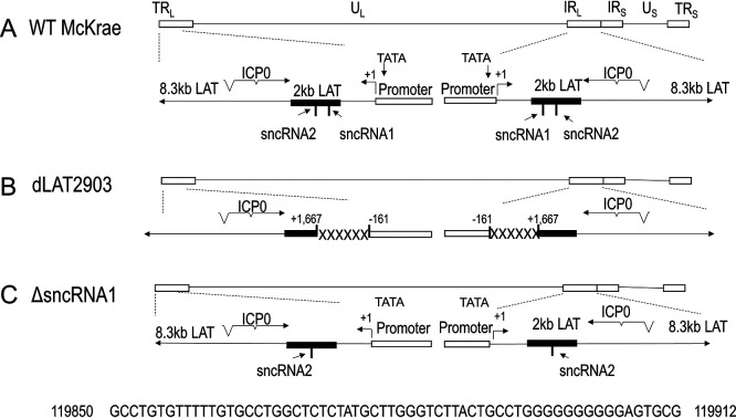
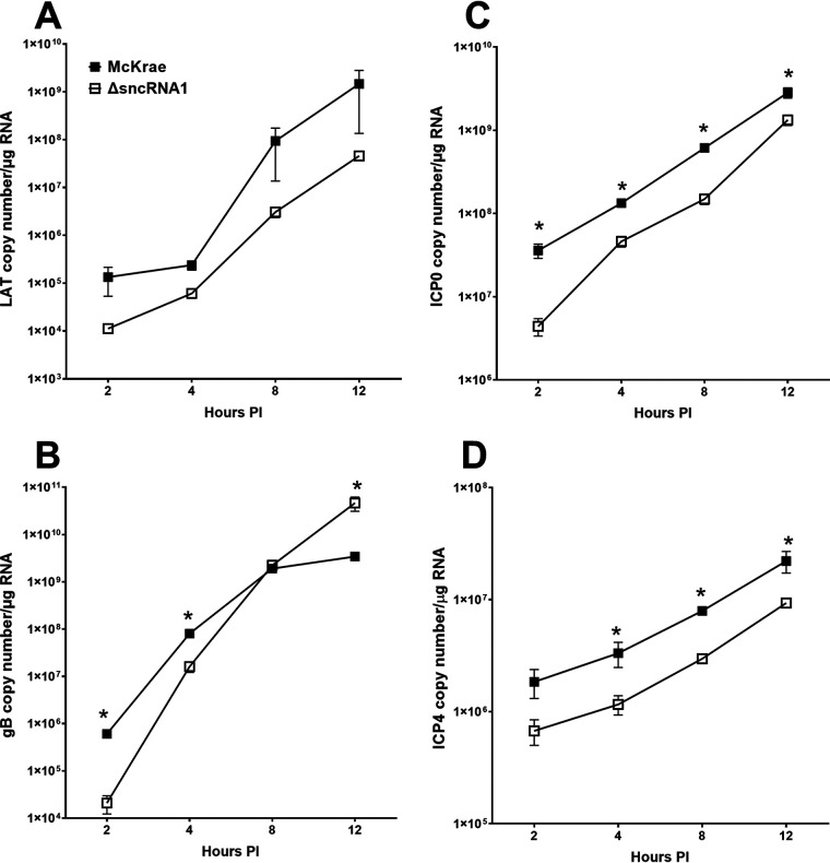
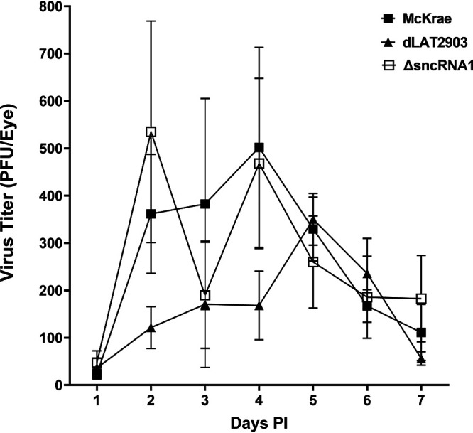
The HSV-1 latency-associated transcript (LAT) locus contains two small noncoding RNA (sncRNA) sequences (sncRNA1 and sncRNA2) that are not microRNAs (miRNAs). We recently reported that sncRNA1 is more important for in vitro activation of the herpesvirus entry mediator than sncRNA2, but its in vivo function is not known. To determine the role, if any, of sncRNA1 during herpes simplex virus 1 (HSV-1) infection in vivo, we deleted the 62-bp sncRNA1 sequence in HSV-1 strain McKrae using dLAT2903 (LAT-minus) virus, creating ΔsncRNA1 recombinant virus. Deletion of the sncRNA1 in ΔsncRNA1 virus was confirmed by complete sequencing of ΔsncRNA1 virus and its parental virus (i.e., McKrae). Replication of ΔsncRNA1 virus in tissue culture or in the eyes of infected mice was similar to that of HSV-1 strain McKrae and dLAT2903 viruses. However, the absence of sncRNA1 significantly reduced the levels of ICP0, ICP4, and gB but not LAT transcripts in infected rabbit skin cells in vitro. In contrast, the absence of sncRNA1 did reduce LAT expression in trigeminal ganglia (TG), but not in corneas, by day 5 postinfection (p.i.) in infected mice. Levels of eye disease in mice infected with ΔsncRNA1 or McKrae virus were similar, and despite reduced LAT levels in TG during acute ΔsncRNA1 infection, McKrae and ΔsncRNA1 viruses did not affect latency or reactivation on day 28 p.i. However, mice infected with ΔsncRNA1 virus were more susceptible to ocular infection than their wild-type (WT) counterparts. Expression of host immune response genes in corneas and TG of infected mice during primary infection showed reduced expression of beta interferon (IFNβ) and IFNγ and altered activation of key innate immune pathways, such as the JAK-STAT pathway in ΔsncRNA1 virus compared with parental WT virus. Our results reveal novel functions for sncRNA1 in upregulating the host immune response and suggest that sncRNA1 has a protective role during primary ocular HSV-1 infection. IMPORTANCE HSV-1 latency-associated transcript (LAT) plays a major role in establishing latency and reactivation; however, the mechanism by which LAT controls these processes is largely unknown. In this study, we sought to establish the role of the small noncoding RNA1 (sncRNA1) encoded within LAT during HSV-1 ocular infection. Our results suggest that sncRNA1 has a protective role during acute ocular infection by modulating the innate immune response to infection.

Keywords: cornea; eye disease; gene expression; immune responses; latency reactivation; recombinant virus; survival; virus replication.


Conflict of interest statement

The authors declare no conflict of interest.

Figures

FIG 1
FIG 1
Construction and structure of the ΔsncRNA1 recombinant virus. (A) Diagram of WT HSV-1 genome showing locations of ICP0, 8.3-kb primary LAT, and 2-kb stable LAT loci in the long terminal repeat and internal long repeat. (B) Diagram of dLAT2903 depicting the removed LAT regions, including a portion of the LAT promoter and downstream sequences of the stable 2-kb LAT. (C) Diagram of ΔsncRNA1 showing restoration of all but the sncRNA1 sequence in the stable 2-kb LAT and its promoter sequences. The missing 62-bp sncRNA1 sequence is shown: TRL, terminal repeat, long; UL; IRL, internal repeat, long; IRS, internal repeat, short; US; TRS, terminal repeat, short.
FIG 2
FIG 2
Loss of sncRNA1 sequence does not affect viral replication in vitro. RS cells were infected with ΔsncRNA1, WT McKrae, or dLAT2903 virus at 0.1 PFU/cell (A), 1 PFU/cell (B), or 10 PFU/cell (C) for 12, 24, or 48 h. Titers of each virus at each time point were determined using a standard plaque assay. No significant differences in titer between the three viruses were seen at any infectious dose or time point. Results are shown as the mean ± SEM (Fisher’s exact test) from four separate experiments (n = 13).
FIG 3
FIG 3
Reduced expression of ICP0, gB, and ICP4 transcripts in ΔsncRNA1-infected cells. RS cells were infected with either WT McKrae or ΔsncRNA1 virus at 1 PFU/cell. Cell lysates were collected at 2, 4, 8, and 12 h p.i., and total RNA was extracted. Relative copy numbers of LAT (A), gB (B), ICP0 (C), and ICP4 (D) were determined by qRT-PCR as described in Materials and Methods. Experiments were repeated twice, and each point represents the mean ± SEM (Student's t test) of results from 10 samples.
FIG 4
FIG 4
Virus titers in tear films of ΔsncRNA1-infected mice are similar to those of McKrae- and dLAT2903-infected mice. Mice were infected ocularly with 2 × 105 PFU/eye of WT McKrae, dLAT2903, or ΔsncRNA1 virus without corneal scarification. Tear films were collected on days 1 to 7 p.i., and virus titers were determined by a standard plaque assay. The experiment was repeated twice, and each point represents the mean ± SEM (Fisher’s exact test) from 20 eyes per group for days 1 to 4, 6, and 7 p.i. and from 40 eyes per group for day 5 p.i.
FIG 5
FIG 5
Reduced LAT expression in TG of mice infected with ΔsncRNA1 virus during primary infection. Mice were ocularly infected with 2 × 105 PFU/eye of WT McKrae or ΔsncRNA1 virus. Ocularly infected mice were euthanized on days 1, 3, and 5 p.i., and corneas and TG were collected. Total RNA was extracted from collected tissues, and relative LAT expression levels in corneas (A) and TG (B) were determined by qRT-PCR as described in Materials and Methods. Each point represents the mean ± SEM (Student's t test) of results from 12 individual corneas or TG from two experiments.
FIG 6
FIG 6
sncRNA1 regulates gene expression of IFNβ1 and IFNγ in TG of infected mice. RNA from day 5 TG samples described in the legend to Fig. 5 were subjected to qRT-PCR, and gene expression levels of IFNα2 (A), IFNβ1 (B), and IFNγ (C) were determined. Fold changes were calculated relative to the level of each transcript in TG of uninfected mice. GAPDH expression was used to normalize the expression of each transcript. Bars represent the mean ± SEM (Student's t test) of results from 12 TG per group in two experiments.
FIG 7
FIG 7
Loss of sncRNA1 sequence affects mouse survival after ocular infection. Mice were ocularly infected with 2 × 105 PFU/eye of McKrae or ΔsncRNA1 virus. Survival of infected mice was monitored for 28 days p.i. The graph represents the average results of two independent experiments with 20 mice per group (chi-square test).
FIG 8
FIG 8
sncRNA1 does not play a role in eye disease. Mice were infected ocularly as described above with HSV-1 strain McKrae or ΔsncRNA1 virus. The extent of corneal scarring (A) and angiogenesis (B) was determined by slit lamp microscopy and scored in a blinded fashion. Bars represent average scores ± SEM of results from two experiments (Student's t test) for 26 and 24 eyes for McKrae- and ΔsncRNA1-infected mice, respectively.
FIG 9
FIG 9
sncRNA1 is not involved in latency or reactivation. Mice were infected ocularly as described above with McKrae or ΔsncRNA1 virus. TG were harvested on day 28 p.i. (A) gB copy number in TG of latently infected mice. DNA isolated from individual TG was used to determine the relative gB copy number by qPCR as described in Materials and Methods. Bars represent the mean gB copy number ± SEM from 7 TG for McKrae-infected mice and from 12 TG from ΔsncRNA1-infected mice (Student's t test). The experiment was repeated twice. (B) LAT copy number in TG of latently infected mice. Isolated RNA from TG was subjected to qRT-PCR, and the LAT RNA copy number was quantified as described in Materials and Methods. Bars represent the mean ± SEM of results from 14 TG per group (Student's t test). The experiment was repeated twice. (C) Explant reactivation in TG of latently infected mice. Mice were infected as described above. On day 28 p.i., individual TG were harvested and the time to reactivation was determined by an explant reactivation assay. Points indicate the day at which CPE was first observed. The horizontal line represents the mean time to reactivation ± SEM from 20 TG for McKrae and 26 TG for ΔsncRNA1 (Student's t test).
FIG 10
FIG 10
Effect of sncRNA1 absence on cellular transcripts in TG of latently infected mice. Mice were infected as described above, and TG were collected from surviving mice on day 28 p.i. Total RNA was isolated, and expression levels of CD8 (A), CD4 (B), CTLA4 (C), Tim-3 (D), PD-1 (E), IFNα2 (F), IFNβ1 (G), and IFNγ (H) were determined using qRT-PCR. Results are presented as the fold increase over baseline mRNA levels in TG of naive mice for each group. GAPDH expression was used to normalize the expression of each transcript. Bars represent the mean ± SEM of results from two experiments: n = 13 for CD8, n = 9 for CD4, n = 8 for CTLA4, n = 4 for Tim-3, n = 15 for PD-1, and n = 12 for IFNα2, IFNβ1, and IFNγ (Student's t test).
FIG 11
FIG 11
Effect of sncRNA1 absence on HVEM gene expression in TG of latently infected mice. Mice were ocularly infected with either WT McKrae, dLAT2903, or ΔsncRNA1 virus as described above. Infected mice were euthanized on day 28 p.i., and total RNA was isolated from individual TG. HVEM expression was determined using qRT-PCR, and fold change was calculated relative to the HVEM levels in TG of uninfected mice. Bars represent the mean ± SEM of results from 9 TG from two independent experiments (Student's t test).
FIG 12
FIG 12
Altered expression of genes involved in T cell activation and apoptosis in ΔsncRNA1-infected mouse corneas. Mice were ocularly infected with 2 × 105 PFU/eye of McKrae or ΔsncRNA1 virus. Corneas were collected on day 3 p.i. (time of peak virus titer in the eye), and total RNA from individual corneas was isolated. RNA samples from two separate experiments and nine mice per group were pooled into three samples for analysis in the NanoString nCounter. The total RNA concentration in each well was 20 ng/μL. Expression levels of the 764 myeloid immune panel genes were analyzed as described in Materials and Methods. Differentially expressed genes in the ΔsncRNA1-infected samples were normalized to housekeeping genes and their corresponding genes in WT McKrae-infected corneas. (A) Heat maps were used to identify genes that were upregulated (green) or downregulated (red) and those displaying no change (black) in mRNA expression. (B) The two upregulated and eight downregulated genes in ΔsncRNA1-infected corneas were further analyzed using Metscape. (C) Based on this analysis, the top 11 pathways were as follows: positive regulation of biological process, metabolic process, immune system process, negative regulation of biological process, response to stimulus, biological adhesion, multicellular organismal process, signaling, multiorganism organism process, developmental process, biological regulation, and regulation of biological process.
FIG 13
FIG 13
Altered expression of genes involved in immune cell activation and apoptosis in ΔsncRNA1-infected mouse TG. Mice were ocularly infected as described in the legend to Fig. 7, and TG were collected on day 5 (peak virus titer in the TG) p.i. The experiment was repeated twice. Total RNA was isolated, and RNA from nine mice were pooled into three samples for analysis in the NanoString nCounter. The total RNA concentration in each well was 20 ng/μL. Expression of 764 myeloid immune panel genes were analyzed as described in Materials and Methods. Differentially expressed genes in the ΔsncRNA1-infected samples were normalized to housekeeping genes and their corresponding genes in WT McKrae-infected TG. (A) Heat maps were used to identify genes that were upregulated (green) or downregulated (red) and those displaying no change (black) in mRNA expression. (B) The five upregulated and seven downregulated genes in ΔsncRNA1-infected TG were further analyzed using Metscape. (C) Based on this analysis, the top pathway was immune system process.

Similar articles

Cited by

References

    1. Koujah L, Suryawanshi RK, Shukla D. 2019. Pathological processes activated by herpes simplex virus-1 (HSV-1) infection in the cornea. Cell Mol Life Sci 76:405–419. 10.1007/s00018-018-2938-1. - DOI - PMC - PubMed
    1. Phelan D, Barrozo ER, Bloom DC. 2017. HSV1 latent transcription and non-coding RNA: a critical retrospective. J Neuroimmunol 308:65–101. 10.1016/j.jneuroim.2017.03.002. - DOI - PubMed
    1. Deshpande S, Banerjee K, Biswas PS, Rouse BT. 2004. Herpetic eye disease: immunopathogenesis and therapeutic measures. Expert Rev Mol Med 6:1–14. 10.1017/S1462399404007604. - DOI - PubMed
    1. Mott KR, Bresee CJ, Allen SJ, BenMohamed L, Wechsler SL, Ghiasi H. 2009. Level of herpes simplex virus type 1 latency correlates with severity of corneal scarring and exhaustion of CD8+ T cells in trigeminal ganglia of latently infected mice. J Virol 83:2246–2254. 10.1128/JVI.02234-08. - DOI - PMC - PubMed
    1. Perng GC, Dunkel EC, Geary PA, Slanina SM, Ghiasi H, Kaiwar R, Nesburn AB, Wechsler SL. 1994. The latency-associated transcript gene of herpes simplex virus type 1 (HSV-1) is required for efficient in vivo spontaneous reactivation of HSV-1 from latency. J Virol 68:8045–8055. 10.1128/jvi.68.12.8045-8055.1994. - DOI - PMC - PubMed

Substances