. 2022 Apr 13;96(7):e0000122.
doi: 10.1128/jvi.00001-22. Epub 2022 Mar 7.

Heat Shock-Binding Protein 21 Regulates the Innate Immune Response to Viral Infection

Affiliations

Heat Shock-Binding Protein 21 Regulates the Innate Immune Response to Viral Infection

Yan Xu et al. J Virol. .

Abstract

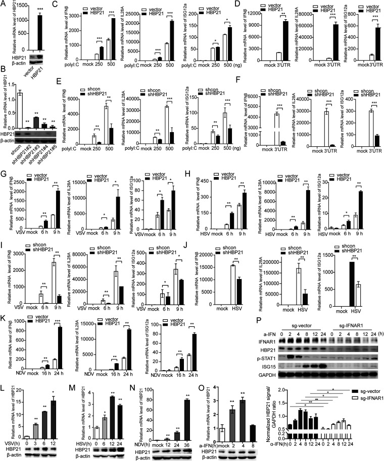
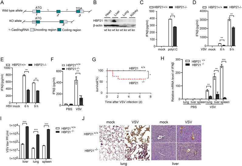
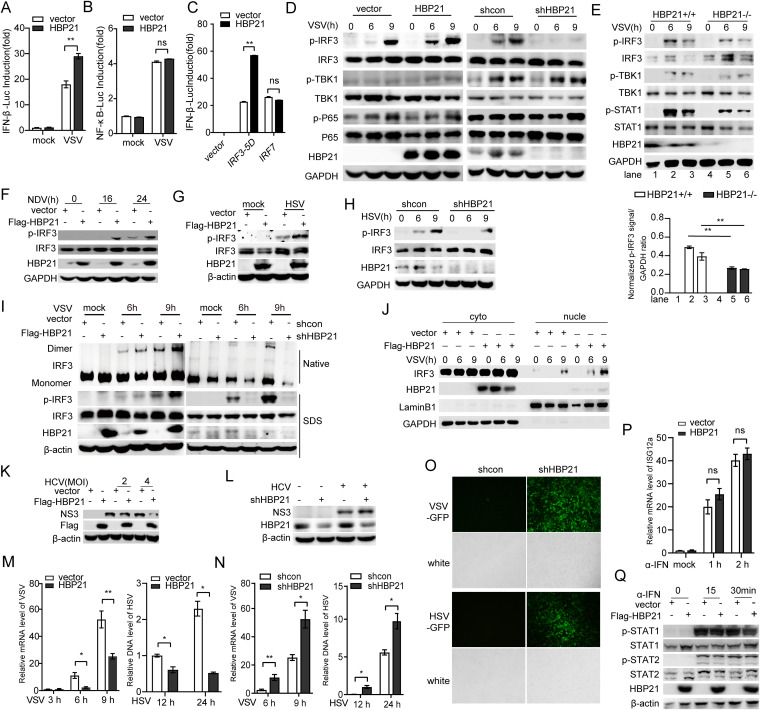
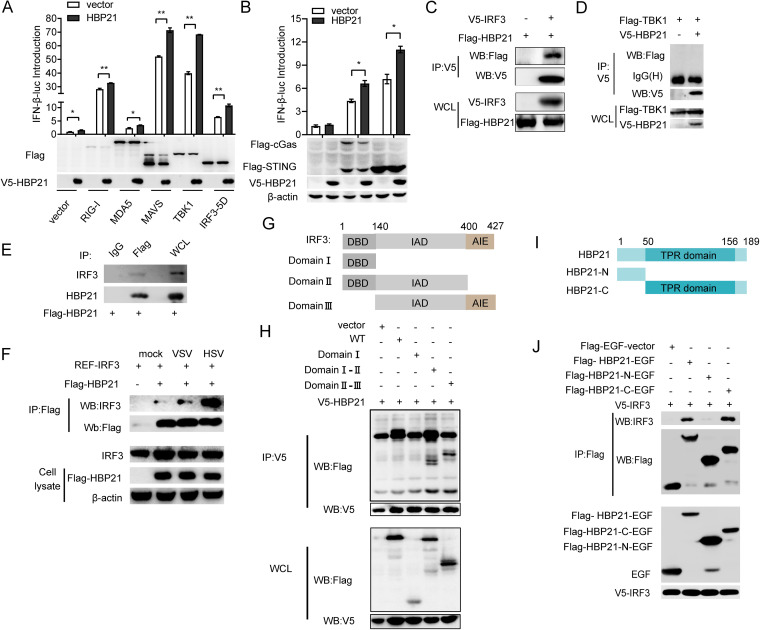
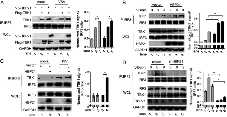
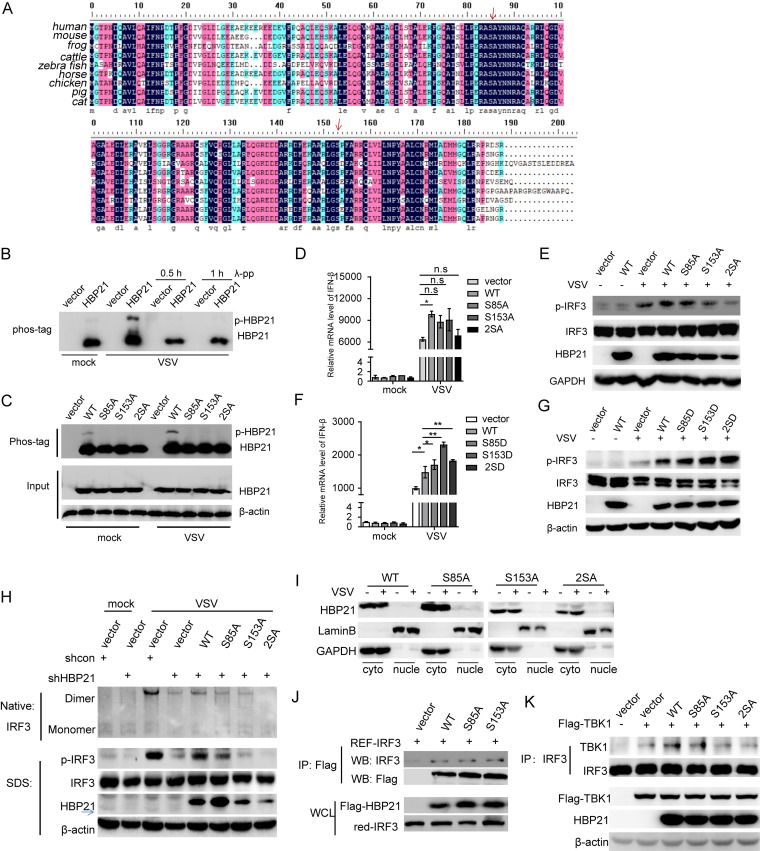
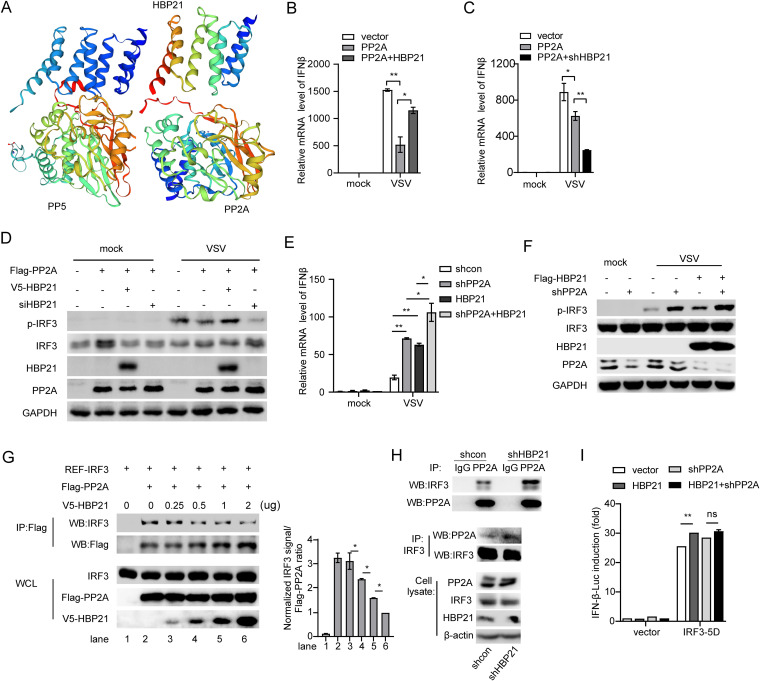
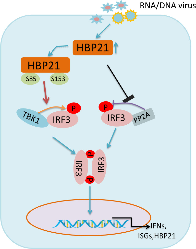
The induction of interferons (IFNs) plays an important role in the elimination of invading pathogens. Heat shock binding protein 21 (HBP21), first known as a molecular chaperone of HSP70, is involved in tumor development. Heat shock binding proteins have been shown to regulate diverse biological processes, such as cell cycle, kinetochore localization, transcription, and cilium formation. Their role in antimicrobial immunity remains unknown. Here, we found that HBP21 drives a positive feedback loop to promote IRF3-mediated IFN production triggered by viral infection. HBP21 deficiency significantly impaired the virus-induced production of IFN and resulted in greater susceptibility to viral infection both in vitro and in vivo. Mechanistically, HBP21 interacted with IRF3 and promoted the formation of a TBK1-IRF3 complex. Moreover, HBP21 abolished the interaction between PP2A and IRF3 to repress the dephosphorylation of IRF3. Analysis of HBP21 protein structure further confirmed that HBP21 promotes the activation of IRF3 by depressing the dephosphorylation of IRF3 by PP2A. Further study demonstrated that virus-induced phosphorylation of Ser85 and Ser153 of HBP21 itself is important for the phosphorylation and dimerization of IRF3. Our study identifies HBP21 as a new positive regulator of innate antiviral response, which adds novel insight into activation of IRF3 controlled by multiple networks that specify behavior of tumors and immunity. IMPORTANCE The innate immune system is the first-line host defense against microbial pathogen invasion. The physiological functions of molecular chaperones, involving cell differentiation, migration, proliferation and inflammation, have been intensively studied. HBP21 as a molecular chaperone is critical for tumor development. Tumor is related to immunity. Whether HBP21 regulates immunity remains unknown. Here, we found that HBP21 promotes innate immunity response by dual regulation of IRF3. HBP21 interacts with IRF3 and promotes the formation of a TBK1-IRF3 complex. Moreover, HBP21 disturbs the interaction between PP2A and IRF3 to depress the dephosphorylation of IRF3. Analysis of HBP21 protein structure confirms that HBP21 promotes the activation of IRF3 by blocking the dephosphorylation of IRF3 by PP2A. Interestingly, virus-induced Ser85 and Ser153 phosphorylation of HBP21 is important for IRF3 activation. Our findings add to the known novel immunological functions of molecular chaperones and provide new insights into the regulation of innate immunity.

Keywords: HSP70; IRF3; PP2A; heat shock-binding protein 21; innate immunity; interferons; molecular chaperone; viral infection.


Conflict of interest statement

The authors declare no conflict of interest.

Figures

FIG 1
FIG 1
HBP21 promotes the innate immune response to viral infection in vitro. (A) Real-time PCR analysis of HBP21 from HLCZ01 cells transfected by p3XFlag-HBP21 was performed, and immunoblotting assays were performed for HBP21 and β-actin antibodies. (B) HLCZ01 cells were transfected with control or specific shRNA targeting HBP21, which was treated with puromycin (2 μg/mL) for 1 month. HBP21 mRNA level was examined by reverse transcription-PCR (RT-PCR) and normalized with GAPDH. Immunoblot analysis of HBP21 and β-actin in the HBP21-silenced HLCZ01 cells was performed. (C to F) HLCZ01 cells transfected with pFlag-HBP21 or empty vector or HBP21-silenced HLCZ01 cells were treated with poly(I·C) at the indicated doses or 200 ng of 3′ UTR for 9 h. The levels of IFN-β, IL-28A, and ISG12a mRNA were examined by RT-PCR and normalized to GAPDH. (G to J) HLCZ01 cells transfected with pFlag-HBP21 or empty vector and HBP21-silenced HLCZ01 cells were infected with VSV (multiplicity of infection MOI = 1) for the indicated times or HSV (MOI = 1) for 9 h. The levels of IFN-β, IL-28A, and ISG12a mRNA were examined by RT-PCR and normalized to GAPDH. (K) Huh7 cells transfected with pFlag-HBP21 or empty vector were infected with NDV (MOI = 0.04) for the indicated times. The levels of IFN-β, IL-28A, and ISG12a mRNA were examined by RT-PCR and normalized to GAPDH. (L to O) Immunoblot analysis and RT-PCR analysis of HBP21 in HLCZ01 cells upon VSV (MOI = 1), HSV (MOI = 0.5), or NDV (MOI = 0.01) infection or with α-IFN (100 U/mL) treatment for the indicated times. (P) Immunoblot analysis of the indicated proteins in HLCZ01-sg-vector and HLCZ01-sg-IFNAR1 stable cell lines treated with IFN-α (100 U/mL) for the indicated times. Values are means and standard deviations (SD). *, P < 0.05; **, P < 0.01; ***, P < 0.001. Data are representative of three experiments.
FIG 2
FIG 2
Pivotal role of HBP21 in the antiviral response in vivo. (A) CRISPR-Cas9 technology was used to edit the HBP21 gene in animal models, and the schematic diagram shows the murine HBP21 allele and the positions of single guide RNA (sgRNA). (B) Immunoblot analysis of HBP21 and β-actin from different tissues to identify the knockout effect. (C) ELISA of IFN-β in the culture medium of HBP21+/+ and HBP21−/− macrophages transfected with 500 ng of poly(I·C) for 6 h. (D and E) ELISA of IFN-β in the supernatant of HBP21+/+ and HBP21−/− macrophages with or without VSV (MOI = 1) or HSV (MOI = 1) infection for the indicated times. (F) ELISA of IFN-β in sera from wild-type (WT) mice (n = 5) and HBP21 knockout mice (n = 5) infected intraperitoneally for 18 h with VSV (1 × 107 PFU/g body weight). Each symbol represents an individual mouse, and small horizontal lines indicate the means. (G) Survival (Kaplan-Meier curve) of wild-type mice (n = 8) and HBP21−/− mice (n = 8) infected intraperitoneally with a high dose of VSV (1.5 × 107 PFU/g body weight) and monitored for 7 days. (H) The level of IFN-β mRNA was quantified in the lungs, livers, and spleens from wild-type mice (n = 3) and HBP21−/− mice (n = 3) after intraperitoneal infection with VSV (1 × 107 PFU/g body weight) for 18 h or not. (I) Plaque assay of VSV in lungs, livers, and spleens of wild-type mice (n = 3) and HBP21−/− mice (n = 3) after intraperitoneal infection with VSV (1 × 107 PFU/g body weight) for 18 h. (J) H&E staining of the lung and liver sections from WT mice or HBP21−/− mice 18 h after infection with VSV (as for panel D). *, P < 0.5, **, P < 0.01; ***, P < 0.001 (unpaired t test [F] or log-rank test [G]; means and SD in panels C, D, E, H, and I). Data are representative of two or three experiments.
FIG 3
FIG 3
HBP21 regulates IRF3-mediated signaling axis and suppresses viral infection. (A) Dual luciferase analysis of the activity of the IFN-β promoter in HEK293T cells cotransfected with encoding HBP21 and IFN-β promoter plasmids upon VSV (MOI = 1) infection for 6 h. (B) Dual luciferase analysis of the activity of NF-κB promoter in HEK293T cells cotransfected with pFlag-HBP21 and NF-κB-luc plasmid upon VSV (MOI = 1) infection for 6 h. (C) Dual luciferase analysis of the activity of the IFN promoter in HEK293T cells cotransfected with pFlag-HBP21, IFN-luc plasmid, and p3XFlag-IRF3-5D or p3XFlag-IRF7. (D) HLCZ01 cells transfected with pFlag-HBP21 or empty vector or HBP21-silenced HLCZ01 cells were infected with VSV (MOI = 1) for the indicated times. Immunoblotting assays were performed with the indicated antibodies. (E) HBP21+/+ and HBP21−/− macrophages were infected with VSV (MOI = 1) for the indicated times. Immunoblotting assays were performed with the indicated antibodies. (F) Huh7 cells transfected with pFlag-HBP21 or empty vector were infected with NDV (MOI = 0.04) for the indicated times. Immunoblotting assays were performed with the indicated antibodies. (G) Immunoblot analysis of p-IRF3 (S396) in HLCZ01 cells transfected with pFlag-HBP21 or empty vector followed with HSV (MOI = 1) infection for 9 h. (H) Immunoblot analysis of p-IRF3 (S396) in HBP21-silenced HLCZ01 or control cells upon HSV (MOI = 1) infection for the indicated times. (I) Immunoblot analyses of IRF3 in dimer or monomer form, IRF3 phosphorylated at Ser396 [p-IRF3(S396)], total IRF3, and HBP21 were performed by native PAGE or SDS-PAGE in HLCZ01 cells transfected with pFlag-HBP21 or HBP21-silenced HLCZ01 cells with VSV infection. (J) Immunoblot analysis of nucleus-cytoplasm extraction of HBP21-overexpressing HLCZ01 cells upon VSV (MOI = 1) infection. (K and L) HLCZ01 cells were transfected with pFlag-HBP21 or empty vector for 24 h and then infected with HCV (MOI = 2 and 4) for 48 h. The shHBP21-stable HLCZ01 or shVector HLCZ01 cells were infected with HCV (MOI = 4) for 72 h. Immunoblot analysis of nonstructural protein 3 (NS3) of HCV was performed. (M and N) Real time-PCR analysis of VSV in HBP21-overexpressing HLCZ01 cells or HBP21-silenced HLCZ01 cells with VSV (MOI = 1) infection. PCR analysis of HSV in HBP21-overexpressing HLCZ01 cells or HBP21-silenced HLCZ01 cells with HSV (MOI = 0.5) infection. (O) The stable HBP21-silenced HLCZ01 cells and control cells were infected for 24 h with VSV-GFP or HSV-GFP (MOI = 0.1). Bars, 100 μm. (P and Q) HLCZ01 cells were transfected with pFlag-HBP21 or empty vector and then treated with IFN-α (100 U/mL) for the indicated times. Real-time PCR analysis of ISG12a. Immunoblot analysis of indicated antibodies. Data are representative of three independent experiments (means and SD are shown in panels A, B, C, M, N, and P).
FIG 4
FIG 4
HBP21 targets and interacts with IRF3. (A) Dual luciferase assay (top) and immunoblot analysis (bottom) of HEK293T cells transfected with the IFN promoter and RIG-I-N, MDA5, MAVS, TBK1, or IRF3-5D and HBP21 or empty vector. (B) HEK293T cells were transfected with the IFN promoter and 0.5 μg STING, 0.5 μg cGAS or 1 μg STING, and HBP21. Dual luciferase assay (top) and immunoblot analysis (bottom) were performed. (C and D) HEK293T cells were cotransfected with pcDNA3.1a-IRF3 and p3XFlag-HBP21 or with p3XFlag-TBK1 and pcDNA3.1a-HBP21, and immunoprecipitation and Western blotting were performed with the indicated antibodies. (E) HEK293T cells were transfected with pFlag-HBP21, and immunoprecipitation and Western blotting were performed with the indicated antibodies. (F) HLCZ01 cells were cotransfected with pREF-IRF3 and pFlag-HBP21, followed by VSV (MOI = 1) or HSV (MOI = 1) infection for 9 h. Then, immunoprecipitation and Western blotting were performed with the indicated antibodies. (G) Schematic illustration of IRF3 truncation. DBD, DNA binding domain; IAD, IRF3 association domain; AIE, C-terminal autoinhibition element. (H) HEK293T cells were cotransfected with the plasmid encoding IRF3 or the indicated domain and HBP21 for 36 h. Co-IP and immunoblotting were performed with the indicated antibodies. (I) Schematic illustration of IRF3 truncation. The carboxyl-terminal region is aa 1 to 50; the N-terminal region is aa 50 to 189. (J) HEK293T cells were cotransfected with plasmid encoding HBP21 or the indicated domain and full-length pcDNA3.1a-IRF3 for 48 h. Co-IP and immunoblotting were performed with the indicated antibodies. Data are representative of three independent experiments.
FIG 5
FIG 5
HBP21 promotes the formation of TBK1-IRF3 complex. (A) HEK293T cells were cotransfected with p3XFlag-TBK1 and pcDNA3.1a-HBP21 for 36 h, followed by VSV infection for 6 h. Immunoprecipitation and Western blotting were performed with the indicated antibodies. (B and C) HLCZ01 cells transfected with p3XFlag-HBP21 were infected with VSV (MOI = 1) or HSV (MOI = 1). Immunoprecipitation was performed with the indicated antibodies. (D) HBP21-silenced HLCZ01 cells were infected with VSV (MOI = 1) for the indicated times. Immunoprecipitation was performed with the indicated antibodies. Data are representative of three independent experiments.
FIG 6
FIG 6
The phosphorylation of HBP21 at Ser85 and Ser153 is important for the activation of IRF3 during viral infection. (A) Alignment of HBP21 homologous protein was produced by the DNAMAN (7.0) program. (B) HEK293T cells were transfected with the plasmid encoding HBP21 for 24 h and then infected with VSV (MOI = 1) for 6 h. The protein was isolated and treated with phosphatase 37°C for 0.5 or 1 h, and immunoblotting of SDS-PAGE (Phos-tag [100 μM], MnCl2 [200 μM]) of HBP21 was performed. (C) Immunoblot analysis of HBP21 in HLCZ01 cells transfected with the plasmid encoding wild-type HBP21 or alanine mutant HBP21 for 24 h, following treatment as for panel B. (D to G) HLCZ01 cells were transfected with the plasmid encoding wild-type HBP21 or alanine or aspartic mutant HBP21 for 36 h and then infected with VSV (MOI = 1) for 6 h. IFN-β mRNA level and phosphorylation of IRF3 at Ser396 were detected by real-time PCR and Western blotting, respectively. (H) HBP21-silenced cells or control cells were transfected by the plasmid encoding wild-type HBP21 or alanine mutant HBP21 and then infected with VSV (MOI = 1) for 9 h. Immunoblot analysis of IRF3 in dimer or monomer form (top) or phosphorylation of IRF3 at Ser396, total IRF3 and HBP21 (bottom) in the cells with VSV infection for 6 and 9 h. (I) HLCZ01 cells transfected with the plasmid encoding wild-type HBP21 or vector alanine mutant HBP21 were infected with VSV (MOI = 1) for 24 h, followed by nucleus-cytoplasm extraction. Immunoblot analysis was performed. (J) HEK293T cells were cotransfected with the plasmid encoding IRF3 and the plasmid encoding wild-type HBP21 or alanine mutant HBP21 for 36 h. Immunoprecipitation and immunoblotting assays were performed with the indicated antibodies Data are representative of three independent experiments. (K) HEK293T cells were cotransfected with the plasmid encoding TBK1 and the plasmid encoding wild-type HBP21 or alanine mutant HBP21 upon VSV infection for 6 h. Immunoprecipitation and Western blotting were performed with the indicated antibodies. Data are representative of three independent experiments (means and SD in panels D and F).
FIG 7
FIG 7
HBP21 blocks the interaction between PP2A and IRF3 to promote the innate immune response to viral infection. (A) The structure analysis of PP5, HBP21, and PP2A was predicted by the online software AlphaFold. (B to D) HLCZ01 cells were cotransfected with the plasmids encoding PP2A and HBP21 or shHBP21, followed by VSV infection for 9 h. IFN-β mRNA and phosphorylation of IRF3 at Ser396 were detected by real-time PCR and Western blotting, respectively. (E and F) HLCZ01 cells were infected with lenti-shPP2A or lenti-shVector and then transfected with the plasmid encoding HBP21, followed by VSV infection for 9 h. IFN-β mRNA and phosphorylation of IRF3 at Ser396 were detected by real-time PCR and Western blotting, respectively. (G) HEK293T cells were cotransfected with the indicated plasmids for 36 h. Immunoprecipitation and Western blotting were performed with the indicated antibodies. (H) HEK293T cells were infected by lenti-shHBP21 or lenti-shVector for 48 h. Immunoprecipitation and Western blotting were performed with the indicated antibodies. (I) Dual luciferase analysis of the activity of the IFN promoter in HEK293T cells infected by lenti-shPP2A or lenti-shVector for 48 h and cotransfected with pV5-HBP21, pIFN-luc, and p3XFlag-IRF3-5D or empty vector. Data are representative of three independent experiments (means and SD in panels B, C, E, and I).
FIG 8
FIG 8
Schematic model of the dual regulation of IRF3 by HBP21. Upon virus infection, HBP21 is induced and phosphorylated at Ser85 and Ser153. The phosphorylated HBP21 at Ser85 and Ser153 enhances the formation of TBK1-IRF3 complex and triggers the phosphorylation of IRF3. Moreover, HBP21 promotes the activation of IRF3 by depressing the dephosphorylation of phosphatase PP2A of IRF3. HBP21 is important for IRF3 dimerization. HBP21 promotes innate immunity and antiviral activity through dual pathways.

Similar articles

Cited by

References

    1. Fensterl V, Chattopadhyay S, Sen GC. 2015. No love lost between viruses and interferons. Annu Rev Virol 2:549–572. 10.1146/annurev-virology-100114-055249. - DOI - PMC - PubMed
    1. Chattopadhyay S, Sen GC. 2017. RIG-I-like receptor-induced IRF3 mediated pathway of apoptosis (RIPA): a new antiviral pathway. Protein Cell 8:165–168. 10.1007/s13238-016-0334-x. - DOI - PMC - PubMed
    1. Loo YM, Gale M, Jr.. 2011. Immune signaling by RIG-I-like receptors. Immunity 34:680–692. 10.1016/j.immuni.2011.05.003. - DOI - PMC - PubMed
    1. King KR, Aguirre AD, Ye YX, Sun Y, Roh JD, Ng RP, Jr, Kohler RH, Arlauckas SP, Iwamoto Y, Savol A, Sadreyev RI, Kelly M, Fitzgibbons TP, Fitzgerald KA, Mitchison T, Libby P, Nahrendorf M, Weissleder R. 2017. IRF3 and type I interferons fuel a fatal response to myocardial infarction. Nat Med 23:1481–1487. 10.1038/nm.4428. - DOI - PMC - PubMed
    1. Mino T, Takeuchi O. 2017. NSD3 keeps IRF3 active. J Exp Med 214:3475–3476. 10.1084/jem.20171980. - DOI - PMC - PubMed

MeSH terms

Substances