

Oxytocin enhances basolateral amygdala activation and functional connectivity while processing emotional faces: preliminary findings in autistic vs non-autistic women
- PMID: 35254443
- PMCID: PMC9527468
- DOI: 10.1093/scan/nsac016
Oxytocin enhances basolateral amygdala activation and functional connectivity while processing emotional faces: preliminary findings in autistic vs non-autistic women
Abstract
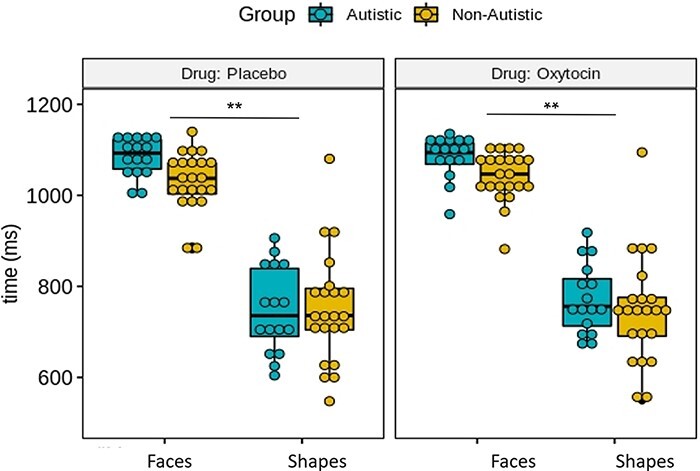
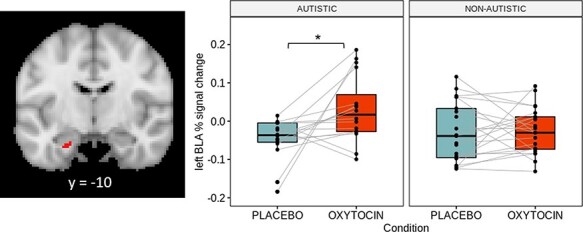
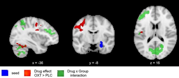
Oxytocin is hypothesized to promote social interactions by enhancing the salience of social stimuli. While previous neuroimaging studies have reported that oxytocin enhances amygdala activation to face stimuli in autistic men, effects in autistic women remain unclear. In this study, the influence of intranasal oxytocin on activation and functional connectivity of the basolateral amygdala-the brain's 'salience detector'-while processing emotional faces vs shapes was tested in 16 autistic and 21 non-autistic women by functional magnetic resonance imaging in a placebo-controlled, within-subject, cross-over design. In the placebo condition, minimal activation differences were observed between autistic and non-autistic women. However, significant drug × group interactions were observed for both basolateral amygdala activation and functional connectivity. Oxytocin increased left basolateral amygdala activation among autistic women (35-voxel cluster, Montreal Neurological Institute (MNI) coordinates of peak voxel = -22 -10 -28; mean change = +0.079%, t = 3.159, PTukey = 0.0166) but not among non-autistic women (mean change = +0.003%, t = 0.153, PTukey = 0.999). Furthermore, oxytocin increased functional connectivity of the right basolateral amygdala with brain regions associated with socio-emotional information processing in autistic women, but not in non-autistic women, attenuating group differences in the placebo condition. Taken together, these findings extend evidence of oxytocin's effects on the amygdala to specifically include autistic women and specify the subregion of the effect.
Keywords: autism; basolateral amygdala; emotional face processing; oxytocin; salience.
© The Author(s) 2022. Published by Oxford University Press.
Figures
Similar articles
-
Oxytocin's effect on resting-state functional connectivity varies by age and sex.Psychoneuroendocrinology. 2016 Jul;69:50-9. doi: 10.1016/j.psyneuen.2016.03.013. Epub 2016 Mar 19. Psychoneuroendocrinology. 2016. PMID: 27032063 Free PMC article.
-
Oxytocin differentially alters resting state functional connectivity between amygdala subregions and emotional control networks: Inverse correlation with depressive traits.Neuroimage. 2017 Apr 1;149:458-467. doi: 10.1016/j.neuroimage.2017.01.078. Epub 2017 Feb 1. Neuroimage. 2017. PMID: 28161309 Clinical Trial.
-
Impact of chronic intranasal oxytocin administration on face expression processing in autistic children: a randomized controlled trial using fMRI.Mol Autism. 2024 Dec 21;15(1):53. doi: 10.1186/s13229-024-00635-z. Mol Autism. 2024. PMID: 39709442 Free PMC article. Clinical Trial.
-
Impact of prosocial neuropeptides on human brain function.Prog Brain Res. 2008;170:463-70. doi: 10.1016/S0079-6123(08)00436-6. Prog Brain Res. 2008. PMID: 18655902 Review.
-
Intranasal oxytocin as strategy for medication-enhanced psychotherapy of PTSD: salience processing and fear inhibition processes.Psychoneuroendocrinology. 2014 Feb;40:242-56. doi: 10.1016/j.psyneuen.2013.11.018. Epub 2013 Dec 1. Psychoneuroendocrinology. 2014. PMID: 24485496 Review.
Cited by
-
Intranasal oxytocin modulates brain activity during emotional processing in children with treatment resistant conduct problems.Sci Rep. 2025 Apr 3;15(1):11422. doi: 10.1038/s41598-025-92276-2. Sci Rep. 2025. PMID: 40180973 Free PMC article. Clinical Trial.
-
The pharmacodynamic modulation effect of oxytocin on resting state functional connectivity network topology.Front Pharmacol. 2025 Jan 6;15:1460513. doi: 10.3389/fphar.2024.1460513. eCollection 2024. Front Pharmacol. 2025. PMID: 39834799 Free PMC article.
-
Connectome dysfunction in patients at clinical high risk for psychosis and modulation by oxytocin.Mol Psychiatry. 2024 May;29(5):1241-1252. doi: 10.1038/s41380-024-02406-x. Epub 2024 Jan 19. Mol Psychiatry. 2024. PMID: 38243074 Free PMC article. Clinical Trial.
-
Autistic and non-autistic individuals show the same amygdala activity during emotional face processing.Mol Autism. 2024 Jan 10;15(1):2. doi: 10.1186/s13229-024-00582-9. Mol Autism. 2024. PMID: 38200601 Free PMC article.
-
Oxytocin effects on amygdala reactivity to angry faces in males and females with antisocial personality disorder.Neuropsychopharmacology. 2023 May;48(6):946-953. doi: 10.1038/s41386-023-01549-9. Epub 2023 Mar 20. Neuropsychopharmacology. 2023. PMID: 36941365 Free PMC article. Clinical Trial.
References
-
- Alaerts K., Bernaerts S., Vanaudenaerde B., Daniels N., Wenderoth N. (2019). Amygdala–hippocampal connectivity is associated with endogenous levels of oxytocin and can be altered by exogenously administered oxytocin in adults with autism. Biological Psychiatry Cognitive Neuroscience and Neuroimaging, 4, 655–63. - PubMed
