Review
. 2022 Mar 7;13(1):39.
doi: 10.1186/s13244-022-01172-0.

Extrapulmonary tuberculosıs: an old but resurgent problem

Affiliations
Review

Extrapulmonary tuberculosıs: an old but resurgent problem

Ali H Baykan et al. Insights Imaging. .

Abstract

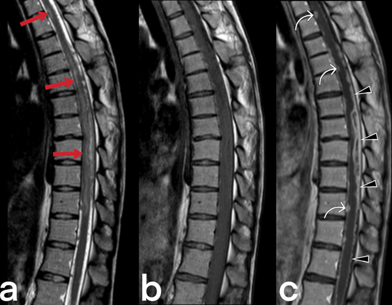
Tuberculosis (TB) primarily affects the lungs, but some of its most devastating clinical consequences arise because of its ability to spread from the lungs to other organs. Extrapulmonary TB (EPTB) constitutes 15-20% of all TB cases. Imaging findings are not always specific and can mimic many diseases; therefore, EPTB should be considered in the differential diagnosis, particularly in patients with immune system disorders (AIDS, patients receiving chemotherapy, etc.) and those in other high-risk groups including people with diabetes. The bacterium's passage to the regional lymph nodes is essential for developing a protective T-cell-mediated immune response, but the bacterium can spread hematologically and via the lymphatic system, leading to extrapulmonary involvement. Diagnosis of EPTB in high-risk patients is made based on suspected clinical and radiological findings, but further positive culture and histopathological confirmation may be required in some instances. Radiological evaluations are critical for diagnosis and crucial in planning the treatment and follow-up. This paper aims to review the typical and atypical imaging features and the differential diagnosis of EPTB.

Keywords: Computed tomography; Extrapulmonary tuberculosis; Magnetic resonance imaging; Tuberculosis; X-ray.


Conflict of interest statement

Mehmet Sukru Erturk is member of the Insights into Imaging Editorial Board. He has not taken part in the review or selection process of this article. The remaining authors declare that they have no competing interests.

Figures

Fig. 1
Fig. 1
Pathogenic life cycle and extrapulmonary dissemination of M. tuberculosis
Fig. 2
Fig. 2
Axial FLAIR (a), pre-contrast (b) axial T1 weighted, and post-contrast (c) axial T1 weighted MR images of a 21-year-old male. Axial FLAIR image shows high signal intensity caused by edema and inflammation in the left frontal lobe (star). In both anterior frontal regions (red arrows) and right parietal area (blue arrows) are seen subdural abscesses. In addition, MR images demonstrate meningeal enhancement and thickening (arrowheads)
Fig. 3
Fig. 3
Radiologic images of a 38-year-old-female who has a diagnosis of miliary TB. Multiple tuberculomas (red arrows) show peripheral rim-like enhancement on post-contrast T1 weighted image in the cerebellum (a). On T2-weighted images (b, c) demonstrate focal lesions (tuberculomas) with central isointense signal and peripheral hyperintensity in gray matter (blue arrows). Multiple nodules secondary to miliary TB are seen (d)
Fig. 4
Fig. 4
MR images of a 63-year-old female with central nervous system TB. Focal lesions (arrows) with rim-like enhancement are seen (tuberculoma) on contrast-enhanced coronal T1 weighted images (a, c). Perilesional increased signals (asterisk and arrowheads) due to vasogenic edema are noted on axial FLAIR images (b, d)
Fig. 5
Fig. 5
MR images of thoracic vertebrae of a 23-year-old female. Sagittal T2-weighted image reveals a heterogeneous signal increase in the spinal cord (red arrows) (a). Precontrast (b) and postcontrast (c) T1 weighted images of the same patient show linear (arrowheads) and nodular (curved arrow) dural enhancement and tuberculous abscess formations in the posterior epidural space
Fig. 6
Fig. 6
MR images of a 56-year-old male with the diagnosis of tuberculous myelitis. Sagittal T2 weighted image (a) shows a heterogeneous increased signal on the spinal cord (arrow). Pre-contrast (b) and post-contrast (c) T1 weighted images reveal linear contrast enhancement of the same region (arrows)
Fig. 7
Fig. 7
MR images of a 17-year-old female with the diagnosis of cervical tuberculous myelitis. Sagittal T2 weighted image (a) shows a heterogeneous increased signal (edema, inflammation) on the spinal cord (arrowheads) and focus (arrow). The cervical spinal cord is observed as an enlarged diffuse hypointense area on pre-contrast T1-weighted image (b). Post-contrast (c) T1 weighted image reveals diffuse heterogeneous and focal (arrow) contrast enhancement of the same region. The focus is a tuberculoma (arrows)
Fig. 8
Fig. 8
A 68-year-old-male. Sagittal reformatted contrast-enhanced CT image reveals an increased thickness of the larynx's posterior wall (arrow). Sternum fracture is also noted in the manubrium
Fig. 9
Fig. 9
Contrast-enhanced T1 weighted coronal (a) and sagittal T2 weighted (b) MR images of a 39-year-old female demonstrate multiple lymphadenopathies in the bilateral cervical chains. The patient was diagnosed with tuberculous lymphadenitis
Fig. 10
Fig. 10
Contrast-enhanced T1 weighted coronal (a) and axial T1 weighted (b) MR images of a 44-year-old female demonstrate multiple lymphadenopathies in the bilateral cervical chains and right supraclavicular region. Multiple intraabdominal lymphadenopathies are also noted (not shown)
Fig. 11
Fig. 11
A 32-year-old male. Painless, discrete, freely mobile, fistulized lumps in the neck are grown progressively (a). Ultrasound showed multiple necrotic lymph nodes (b). Scrofula of the neck
Fig. 12
Fig. 12
A 50-year-old female. Coronal pre-contrast (a) and post-contrast (b) T1 weighted images reveal enlarged palatine tonsils, which show peripheral enhancement (stars)
Fig. 13
Fig. 13
A 19-year-old male patient. T2 weighted coronal (a), axial FLAIR (b), and T1 weighted post-contrast axial (c) images show periorbital involvement (arrowheads)
Fig. 14
Fig. 14
A 28-year-old female patient. T1 weighted post-contrast image (a) and T2 weighted image (b) with a tuberculous abscess in the left lacrimal sac (arrows)
Fig. 15
Fig. 15
Axial contrast-enhanced CT (a), axial T2 weighted (b), pre (c), and post-contrast T1 (d) weighted images of a 69-year-old female. Biloculated abscess formation is seen in the anterior left paramedian region of the neck, which shows peripheral enhancement
Fig. 16
Fig. 16
Contrast-enhanced thorax CT image of a 70-year-old female. Multiple mediastinal lymphadenopathies are noted in the prevascular region (red arrows)
Fig. 17
Fig. 17
The chest X-ray (a) and coronal reformatted CT (b) image without contrast of a 4-year-old girl demonstrate unilateral pleural effusion in the right side (stars). Tuberculous pleurisy
Fig. 18
Fig. 18
Transverse (a) and coronal reformatted (b) CT images without contrast of a 24-year-old female. A mass-like focal pleural lesion in the right side containing a few coarse calcifications is noted. The patient is diagnosed with TB on histopathologic assessment after surgical removal of the mass
Fig. 19
Fig. 19
Transverse (a) and sagittal reformatted (b) CT images without intravenous contrast of a 55-year-old male. Pericardial thickening with linear calcification (arrowheads), right-sided pleural effusion (black asterisk), and infected fluid collection (white asterisk) in the left pleural cavity are seen. Mediastinal and pericardial tuberculous
Fig. 20
Fig. 20
Ultrasound images from four different female patients (ad). Abscesses (arrowheads), sinus tracts (arrows), and hypoechoic parenchymal edema areas (stars)
Fig. 21
Fig. 21
MR images of a 41-year-old female. Axial T2 weighted (a, d), pre-contrast axial T1 weighted (b, e), and post-contrast axial T1 weighted (c, f) images reveal diffuse increased signal intensity, cystic lesions (arrowheads), ductal ectasia (arrow), and thickened skin (curved arrow) in the right breast. Diffuse-type breast TB
Fig. 22
Fig. 22
Breast MR images of a 39-year-old female. T2 weighted image (a), T1 weighted image (b) post-contrast (c), and subtracted image (d) demonstrate thickening of the skin (arrowhead) and multiple abscesses (arrows) with peripheral enhancement in the right breast
Fig. 23
Fig. 23
Transverse abdomen CT image (a) of a 17-year-old female with known TB. Coarse calcifications in the liver parenchyma suggestive of TB sequela are noted. Coronal reformatted CT image of the same patient (b) reveals multiple lymphadenopathies with peripheral calcifications
Fig. 24
Fig. 24
A 19-year-old male. Post-contrast CT image shows multiple mesenteric lymphadenopathies with peripheral enhancement
Fig. 25
Fig. 25
Transverse CT image with intravenous contrast of a 29-year-old male. Massive ascites is indicated in the abdomen cavity (stars). The peritoneum's thin linear contrast enhancement is also noted. Tuberculous peritonitis
Fig. 26
Fig. 26
A 24-year-old male patient. Axial (a) and coronal postcontrast CT (b) images show mesenteric striation (arrowheads) and ascites (star)
Fig. 27
Fig. 27
A 33-year-old female. Axial post-contrast CT images (a, b) demonstrate omental nodularity, mesenteric fat stranding (arrowheads), and ascites (star). Intraabdominal lymphadenopathies are also evident (arrows) in image b
Fig. 28
Fig. 28
Axial contrast-enhanced CT (a) and axial T2 weighted MR (b) images of a 66-year-old female demonstrate multiple foci (arrowheads) in the spleen. Note small hypodense areas with peripheral enhancement in foci (a). Splenic TB
Fig. 29
Fig. 29
A coronal contrast-enhanced CT image of a 43-year-old male shows multiple hypodense lesions (arrows) in the liver and spleen. Hepatic and splenic TB
Fig. 30
Fig. 30
Abdominal CT scan and MR images of the same patient (in Fig. 27). Contrast-enhanced CT (a) and T1 weighted contrast-enhanced MR images (b) show a small abscess formation with rim-like enhancement in the anterior subcapsular region of the liver. Diffusion-weighted images (DWI) (c) and apparent diffusion coefficient (ADC) map (d) reveal diffusion restriction consistent with an abscess
Fig. 31
Fig. 31
A 35-year-old male. Magnetic resonance cholangiopancreatography (MRCP) images (a, b) show extensive multifocal intrahepatic biliary strictures (arrows) and dilated common hepatic duct (star)
Fig. 32
Fig. 32
A 40-year-old female. Contrast-enhancement CT scan (a, b) and Doppler ultrasonography (c) demonstrated a large heterogeneous cystic-necrotic mass in the head of the pancreas
Fig. 33
Fig. 33
Transverse CT image without intravenous contrast of a 24-year-old female. Diffuse-symmetric wall thickening of the ileal segment is noted (arrows). Ileal TB
Fig. 34
Fig. 34
A 31-year-old female. Contrast-enhanced CT image demonstrates diffuse-symmetric wall thickening and enhancement of the cecum with surrounding inflammatory changes (arrows)
Fig. 35
Fig. 35
A 52-year-old female. Contrast-enhancement CT (a), T2 weighted MR (b), and contrast-enhancement T1 weighted MR (c) images demonstrate a heterogeneous lesion with peripheral enhancement (arrows) in the right adrenal gland (adrenal TB). Multiple calcified heterogeneous areas (arrowheads) are also seen in the liver (hepatic TB)
Fig. 36
Fig. 36
Contrast-enhancement CT scan of a 48-year-old female with renal tuberculous show debris collection within dilated calyces and parenchymal destruction (with cortical thinning)
Fig. 37
Fig. 37
Axial (a) and coronal (b) non-contrast CT scan of a 74-year-old male with urinary tuberculous reveal debris collection within the dilated right renal pelvis and dilated right ureter with diffuse wall thickening. 3D MIP Coronal T2-weighted MR urogram shows strictures (arrows) of the right ureter
Fig. 38
Fig. 38
A 47-year-old male. Coronal post-contrast CT scan shows diffuse thickening of the bladder wall (arrow) and dilated left proximal ureter (arrowheads). Urinary TB
Fig. 39
Fig. 39
Tuberculous prostatitis (abscesses) in a 53-year-old male. T2 weighted axial (a) and sagittal (b) MR images demonstrate focal, hypointense lesions within the prostate gland (arrows). Contrast-enhancement T1 weighted axial (c) and sagittal (b) MR images show heterogeneous lesions with peripheral enhancement (arrows). A rectal tumor (star) is also seen
Fig. 40
Fig. 40
A 38-year-old male. Scrotal US image (a) and post-contrast coronal T1 weighted MR image (b) reveal diffuse heterogeneity, deformation, and dilated cystic lump with peripheral enhancement in the right epididymis (arrows)
Fig. 41
Fig. 41
Bilateral tubo-ovarian TB in a 15-year-old-female with pelvic pain. Axial pre-contrast T1 weighted (a), post-contrast T1 weighted (b), and T2 weighted (c) MR images show bilaterally enlarged ovary (stars) and dilated fallopian tubes (arrowheads). On T2-weighted MR image obtained after antituberculous therapy of 10 months, bilateral ovaries (arrows) and fallopian tubes (not shown) appear normal (d)
Fig. 42
Fig. 42
Bilaterally hydrosalpinx (right: blue arrows, left: red arrow) and ascites (stars) in the peritoneal cavity are observed on coronal contrast-enhancement CT images (a, b) of the same patient (in Fig. 41)
Fig. 43
Fig. 43
Sagittal-reformatted abdomen CT image of a 22-year-old female with lower abdominal pain and fever obtained after C/S surgery. The uterus is enlarged due to prior pregnancy. Free fluid is noted in the pelvis (arrows). Tuberculous pelvic inflammatory disease
Fig. 44
Fig. 44
Contrast-enhanced T1 weighted axial MR image of a 52-year-old male appears an inflammatory mass lesion within the adjacent soft tissues to the body of the fourth lumbar vertebra be caused by a phlegmon and abscess
Fig. 45
Fig. 45
MR images of lumbar vertebrae of a 47-year-old male with tuberculous spondylodiscitis. Sagittal T2-weighted image (a) shows the heterogeneous low signal in the anterior parts of the L3 and L4 vertebral bodies and L3-4 intervertebral disc, which offers minimal anterior subligamentous extension (arrows). Pre-contrast (a) and post-contrast (b) T1 weighted images reveal diffuse contrast enhancement of both vertebral bodies in keeping with spondylodiscitis (arrowheads)
Fig. 46
Fig. 46
A 41-year-old male with pain in the posterior neck region. Non-contrast axial CT image (a) demonstrates destruction and bone erosion of the posterior spinous process of the C7 vertebra (arrowheads). Pre-contrast sagittal T1 weighted image (b) reveals diffuse decreased signal intensity within the c7 vertebral body and posterior spinous process. The lesion shows prominent diffuse contrast enhancement on sagittal (c) T1-weighted images (stars)
Fig. 47
Fig. 47
Pelvic x-ray of a 46-year-old male. The late finding of tuberculous sacroiliitis is characterized by degenerative changes in the left sacroiliac joint (arrows)
Fig. 48
Fig. 48
MR images of a 43-year-old male. Pre-contrast T1 weighted (a) and post-contrast (b) T1 weighted images demonstrate contrast enhancement in the synovium of the left sacroiliac joint in keeping with acute sacroiliitis (arrows). Arrowhead shows an abscess formation in the left iliac muscle that enhances peripherally after contrast injection
Fig. 49
Fig. 49
Coronal (a) and sagittal (b) reformatted CT images of a 70-year-old female who has a complaint of swelling in the anterior wall of the chest. Both images reveal mass-like lesions in the right sternoclavicular joint, causing bone destruction containing small calcified areas. The patient was diagnosed with TB after the biopsy
Fig. 50
Fig. 50
Pelvic X-ray of a 53-year-old female. The left hip joint sequela of TB is characterized by degenerative arthritis (arrows)
Fig. 51
Fig. 51
CT (a) and lateral ankle direct X-ray (b) images of two different patients. Lytic areas are observed in the tibia in both cases with proven tuberculous osteomyelitis and abscess
Fig. 52
Fig. 52
MR images of a 59-year-old male. Pre-contrast (a) and postcontrast (b) T1 weighted images demonstrate intraosseous (arrowheads) and soft tissue abscesses (arrow) in the distal part of the tibia and plantar face of the foot
Fig. 53
Fig. 53
Sagittal reformatted CT (a) and MR images (bd) of a 48-year-old male. CT image shows focal low-attenuation areas in the distal part of the tibia in keeping with abscesses and bone destructions (arrowheads). On sagittal T2-weighted (b), pre-contrast T1-weighted (c), and post-contrast T1 weighted (d) MR images reveal extensive signal intensity change within the bone marrow of the talus and distal tibial part (stars). MR images also demonstrate an abscess in the adjacent soft tissue to the Achilles tendon (arrows)
Fig. 54
Fig. 54
MR images of a 60-year-old male. Pre-contrast axial (a), post-contrast axial (b), and sagittal (c) T1 weighted images reveal intraosseous abscesses (arrowheads) with peripheral enhancement in the distal part of the femur and proximal tibia
Fig. 55
Fig. 55
A contrast-enhanced CT image of a 69-year-old male demonstrates the left iliopsoas abscess (arrows)
Fig. 56
Fig. 56
T2 weighted (a) and post-contrast T1 weighted MR images of a 35-year-old male demonstrate abscess formation with peripheral enhancement in the left iliac muscle (b)

References

    1. Heye T, Stoijkovic M, Kauczor H, Junghanss T, Hosch W. Extrapulmonary tuberculosis: radiological imaging of an almost forgotten transformation artist. RoFo. 2011;183(11):1019–1029. - PubMed
    1. World Health Organization. Global tuberculosis report. 2021. https://www.who.int/teams/global-tuberculosis-programme/tb-reports/globa.... Accessed 14 Oct 2021.
    1. Gambhir S, Ravina M, Rangan K, Dixit M, Barai S, Bomanji J. Imaging in extrapulmonary tuberculosis. Int J Infect Dis. 2017;56:237–247. - PubMed
    1. Sharma SK, Mohan A, Kohli M (2021) Extrapulmonary tuberculosis. Expert Rev Respir Med 15(7):931–948 - PubMed
    1. Sharma SK, Mohan A. Extrapulmonary tuberculosis. Indian J Med Res. 2004;120(4):316–353. - PubMed

LinkOut - more resources