. 2022 Mar 15;119(11):e2115480119.
doi: 10.1073/pnas.2115480119. Epub 2022 Mar 7.

Accurate positioning of functional residues with robotics-inspired computational protein design

Affiliations

Accurate positioning of functional residues with robotics-inspired computational protein design

Cody Krivacic et al. Proc Natl Acad Sci U S A. .

Abstract

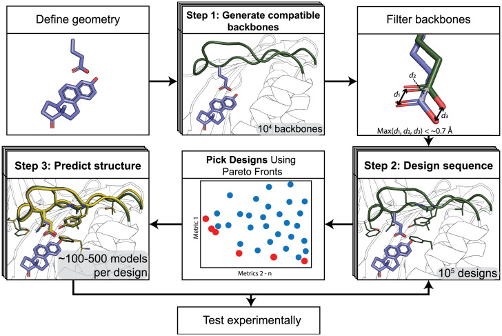
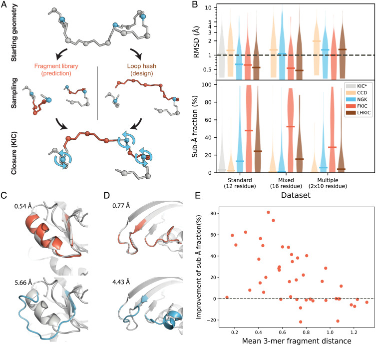
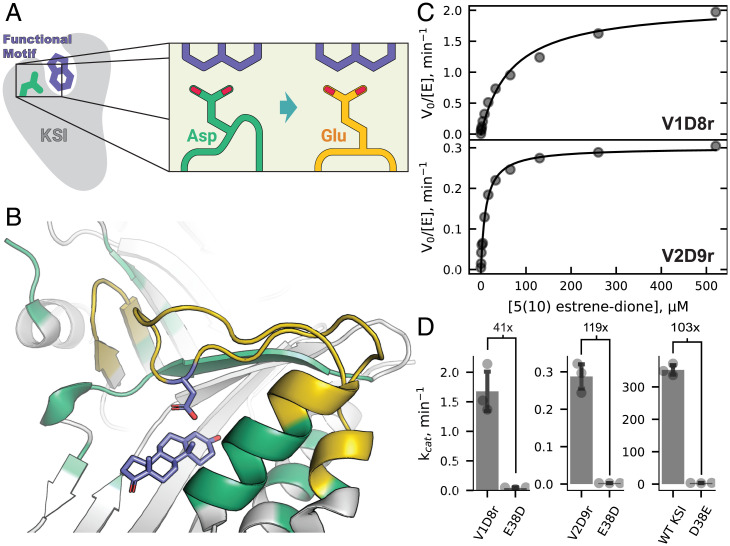
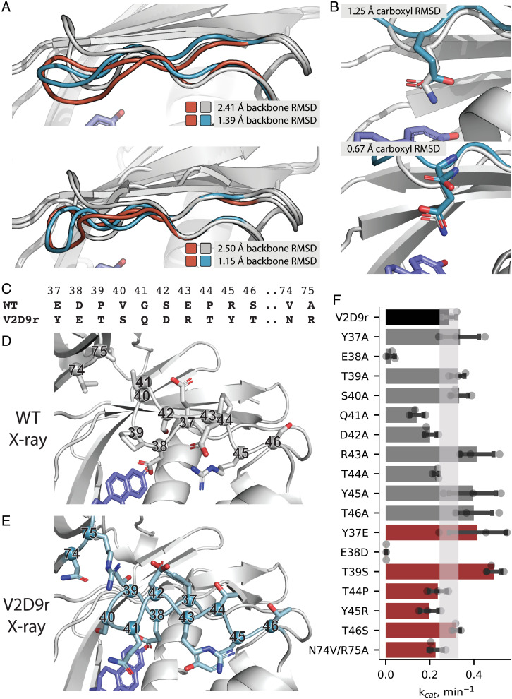
SignificanceComputational protein design promises to advance applications in medicine and biotechnology by creating proteins with many new and useful functions. However, new functions require the design of specific and often irregular atom-level geometries, which remains a major challenge. Here, we develop computational methods that design and predict local protein geometries with greater accuracy than existing methods. Then, as a proof of concept, we leverage these methods to design new protein conformations in the enzyme ketosteroid isomerase that change the protein's preference for a key functional residue. Our computational methods are openly accessible and can be applied to the design of other intricate geometries customized for new user-defined protein functions.

Keywords: Rosetta; computational protein design; design of function; structure prediction.


Conflict of interest statement

Competing interest statement: J.J.G. and T.K. are unpaid board members of the Rosetta Commons.

Figures

Fig. 1.
Fig. 1.
Steps of the PIP protocol. Top Left: functional geometry is defined. Top Middle: New backbone conformations (green) are generated to satisfy the geometric restraints. Top Right: Backbones are filtered based on their ability to satisfy the geometric restraints. d1, d2, and d3 refer to the distances of the atoms in a positioned carboxyl group to their defined ideal positions. Bottom Right: Sequences are designed to stabilize the de novo backbone. Bottom Middle: Designs are selected based on multiple computational quality metrics using Pareto fronts (SI Appendix, SI Methods). Red: Pareto-efficient designs; blue: other designs. Bottom Left: For selected sequences, Rosetta structure prediction method are applied to predict the lowest-energy structure (yellow). Illustrations use the KSI model system detailed in Fig. 3.
Fig. 2.
Fig. 2.
FKIC improves prediction of conformations of local backbone segments. (A) Individual FKIC/LHKIC move. Three Cα atoms (blue) on the target segment to be modeled (gray) are picked randomly as pivots. Fragment insertion (FKIC) or loop hash (LHKIC) is applied to sample torsion degrees of freedom at nonpivot atoms (red), which breaks the chain. The KIC algorithm is then used to close the chain by determining appropriate values for the pivot torsions. (B) Comparison of performance of different methods for three datasets: 1) the Standard dataset described in ref. and two new sets, 2) a “Mixed Segment” dataset with 30 16-residue regions that contain both loops and segments of regular secondary structure and 3) a “Multiple Segments'' dataset of 30 cases with two separate 10-residue regions that are interacting. KIC (17): gray; CCD (24): orange; NGK (26): blue; FKIC: red; LHKIC: brown. Upper: violin plot of RMSD of lowest energy (best) model across each dataset. Horizontal bars indicate the median lowest-energy RMSD. FKIC is the only method that provides predictions with atomic accuracy (≤1 Å median RMSD) for all datasets. Lower: violin plot of fraction of predicted models in each dataset that have subångstrom accuracy. FKIC leads to considerable improvements over previous methods. Asterisk indicates data from ref. ; all other simulations were run with the ref2015 Rosetta energy function (21); methods using fragments (CCD and FKIC) used identical fragment libraries that excluded fragments from structural homologs to the target proteins. (C and D) FKIC accurately predicts geometries from sequence in which the previous state-of-the-art method, NGK, fails. Shown are examples from the Mixed Segment (C) and Multiple Segments dataset (D). Experimentally determined structures: gray; predictions from FKIC: red, Top; predictions from NGK: blue, Bottom. RMSDs to the experimentally determined structures are given in each panel in Å. (E) The fraction of subångstrom predictions is negatively correlated with the mean 3-mer fragment distance (Methods). Each data point represents a protein from the Standard 12-residue dataset.
Fig. 3.
Fig. 3.
Functional characterization of designs V1D8r and V2D9r. (A) Schematic of design goal for KSI. Green: wild-type KSI with catalytic aspartate. Yellow: Designed KSI variant with reshaped active site to position the glutamate carboxyl group in place of the wild-type aspartate carboxyl group. (B) KSI wild-type structure (PDB 1QJG), showing the active site regions to be remodeled. Residues allowed to change identity (design) or conformation (repack) during the design process (PIP version 2) are shown in yellow or green, respectively, and static positions are shown in gray. (C) Representative Michaelis-Menten curves for design V1D8r (Top) or V2D9r (Bottom). (D) Bar plots showing the kcat values of V1D8r (Top), V2D9r (Middle), or wild-type KSI (Bottom) and their E38D or D38E active site mutations. Values show the fold change in kcat between the respective D/E active site residue pairs. SDs of independent triplicate experiments are shown as error bars, with individual measurements shown as points.
Fig. 4.
Fig. 4.
Structural characterization of designs V1D8r and V2D9r. (A) Overlay of wild-type KSI crystal structure (gray), lowest-energy predicted models for V1D8r (orange, Top) and V2D9r (orange, Bottom), and crystal structures for V1D8r (blue, Top) and V2D9r (blue, Bottom). (B) Crystal structure (blue) of V1D8r (Top) and V2D9r (Bottom) showing the catalytic glutamate’s placement relative to the amide in the KSI starting structure (PDB 1QJG) used to define the catalytic position (gray). RMSD values between compared structures are indicated in the different panels. (CF) Mutational analysis of differences between wild-type KSI and design V2D9r: sequence alignment (C), comparison between the active site region in the crystal structures of wild-type KSI (D) and in design V2D9r (E), and (F) bar graph of kcat values for design V2D9r (black), alanine scan mutants (gray), and reversion/selected mutants (red). In F, SDs of independent triplicate experiments are shown as error bars with individual measurements shown as points. The kcat error range for V2D9r is shown as a shaded bar.

References

    1. Kuhlman B., Bradley P., Advances in protein structure prediction and design. Nat. Rev. Mol. Cell Biol. 20, 681–697 (2019). - PMC - PubMed
    1. Pan X., Kortemme T., Recent advances in de novo protein design: Principles, methods, and applications. J. Biol. Chem. 296, 100558 (2021). - PMC - PubMed
    1. Glasgow A. A., et al. , Computational design of a modular protein sense-response system. Science 366, 1024–1028 (2019). - PMC - PubMed
    1. Marcandalli J., et al. , Induction of potent neutralizing antibody responses by a designed protein nanoparticle vaccine for respiratory syncytial virus. Cell 176, 1420–1431.e17 (2019). - PMC - PubMed
    1. Silva D. A., et al. , De novo design of potent and selective mimics of IL-2 and IL-15. Nature 565, 186–191 (2019). - PMC - PubMed