. 2022 Mar 15;119(11):e2118930119.
doi: 10.1073/pnas.2118930119. Epub 2022 Mar 7.

Restriction of viral gene expression and replication prevents immortalization of human keratinocytes by a beta-human papillomavirus

Affiliations

Restriction of viral gene expression and replication prevents immortalization of human keratinocytes by a beta-human papillomavirus

Tina M Rehm et al. Proc Natl Acad Sci U S A. .

Abstract

SignificanceHigh-risk (HR) human papillomaviruses (HPV) from the genus alpha cause anogenital and oropharyngeal cancers, whereas the contribution of HPV from the genus beta to the development of cutaneous squamous cell cancer is still under debate. HR-HPV genomes display potent immortalizing activity in human keratinocytes, the natural target cell for HPV. This paper shows that immortalization of keratinocytes by the beta-HPV49 genome requires the inactivation of the viral E8^E2 repressor protein and the presence of the E6 and E7 oncoproteins but also of the E1 and E2 replication proteins. This reveals important differences in the carcinogenic properties of HR-HPV and beta-HPV but also warrants further investigations on the distribution and mutation frequencies of beta-HPV in human cancers.

Keywords: E8^E2; beta-human papillomavirus; cutaneous squamous cell cancer; immortalization.


Conflict of interest statement

The authors declare no competing interest.

Figures

Fig. 1.
Fig. 1.
(A) Linear representation of the HPV49 genome. The URR, early genes (E1 to E8) and the late genes L1 and L2 are indicated. Nucleotide positions of identified SD (D) and SA (A) sites are given. Spliced transcripts identified by qPCR and sequencing are indicated by gray bars, and the putative extension of these transcripts to the potential early polyadenylation site is indicated by dashed lines. (B) qPCR analyses of spliced HPV49 transcripts expressed 3, 6, and 9 d p.t. of NHK. PGK1 was used as a reference transcript. The data are derived from four independent experiments using cells from different donors.
Fig. 2.
Fig. 2.
(A) qPCR analyses of spliced HPV49 transcripts expressed 3, 6, and 9 d p.t. of NHK with HPV49 wt, E1−, E2−, E6−, E7−, or E8− genomes. PGK1 was used as a reference transcript. The data are derived from four independent experiments using cells from different donors. The error bars indicate the SEM. Statistical significance was determined using a ratio-paired t test using the wt as a reference (*P < 0.05; **P < 0.01). (B) qPCR analyses of E6 and E7 transcripts in polyA-enriched RNA in NHK transfected with wt or E8− genomes. The error bars indicate the SEM. Statistical significance was determined using a ratio-paired t test using the wt as a reference (*P < 0.05; **P < 0.01) (C) Southern blot analysis of low–molecular-weight DNA isolated 6 d p.t. of NHK transiently transfected with HPV49 wt and E8− genomes. DNA was digested with DpnI to remove nonreplicated input DNA and EheI to linearize the viral genomes. As a marker (M), 100 pg linearized HPV49 genome was used.
Fig. 3.
Fig. 3.
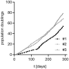
Growth curves of HPV49 E8−-positive human keratinocyte cell lines. Cell lines were continuously cultivated for 300 d and growth was recorded.
Fig. 4.
Fig. 4.
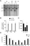
(A) Southern blot analyses of total cellular DNA isolated from stable HPV49 E8− cell lines No. 1 to 3 using 32P-labeled HPV49 genomes. DNA was digested with NotI (N; a noncutter of HPV49) or SalI (S; a single-cutter of HPV49). The positions of concatemeric/integrated (c/i), open circle (oc), linearized (lin), and closed covalent circle (ccc) forms of the viral DNA are indicated. As a marker (M), 100 pg linearized HPV49 genome was used. (B) Total cellular DNA was incubated with or without exonuclease V, and then the presence of ACTB or HPV49 was determined by qPCR. The data are shown as the fraction resistant to exonuclease V digest. The data are derived from three to four independent DNA preparations. The error bars indicate the SEM. (C) HPV49 genome copy numbers in cell lines No. 1 to 3 were determined in total cellular DNA by qPCR using standard curves for HPV49 and ACTB and are given as viral copies per cell assuming two ACTB copies per cell. The error bars indicate the SEM. (D) qPCR analyses of different spliced HPV49 transcripts expressed in cell lines No. 1 to 3 in early and late passage cells. PGK1 was used as a reference transcript. The error bars indicate the SEM.
Fig. 5.
Fig. 5.
QPCR analyses of HPV49 transcripts expressed 6 d p.t. of NHK with HPV49 E8−, E8−/E1−, E8−/E2−, E8−/E6−, or E8−/E7− genomes. PGK1 was used as a reference transcript. The data are derived from four independent transfections using cells from different donors. The error bars indicate the SEM. Statistical significance was determined using a ratio-paired t test using wt as a reference (*P < 0.05; **P < 0.01).
Fig. 6.
Fig. 6.
HPV49 E8− cell lines were transfected with different siRNAs (siControl, si49E6-1, and si49E6-2). The amounts of E6 and E7 transcripts in total RNA were measured by qPCR using PGK1 as a reference (A), and cell numbers (B) were determined 3 d p.t. The error bars indicate the SEM. Statistical significance was determined using a one-sample t test using siControl-transfected cells as a reference (*P < 0.05; **P < 0.01; ***P < 0.001).

References

    1. McLaughlin-Drubin M. E., Munger K., Viruses associated with human cancer. Biochim. Biophys. Acta 1782, 127–150 (2008). - PMC - PubMed
    1. de Martel C., Plummer M., Vignat J., Franceschi S., Worldwide burden of cancer attributable to HPV by site, country and HPV type. Int. J. Cancer 141, 664–670 (2017). - PMC - PubMed
    1. Schiller J. T., Lowy D. R., Understanding and learning from the success of prophylactic human papillomavirus vaccines. Nat. Rev. Microbiol. 10, 681–692 (2012). - PMC - PubMed
    1. Bandolin L., et al. , Beta human papillomaviruses infection and skin carcinogenesis. Rev. Med. Virol. 30, e2104 (2020). - PubMed
    1. Howley P. M., Pfister H. J., Beta genus papillomaviruses and skin cancer. Virology 479–480, 290–296 (2015). - PMC - PubMed

LinkOut - more resources