. 2022 Mar 15;119(11):e2121353119.
doi: 10.1073/pnas.2121353119. Epub 2022 Mar 7.

Structural basis for the oligomerization-mediated regulation of NLRP3 inflammasome activation

Affiliations

Structural basis for the oligomerization-mediated regulation of NLRP3 inflammasome activation

Umeharu Ohto et al. Proc Natl Acad Sci U S A. .

Erratum in

Abstract

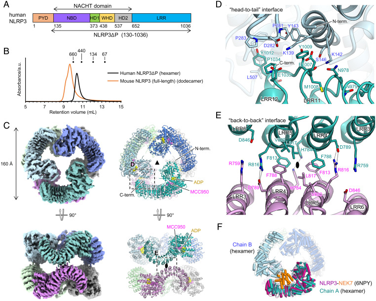
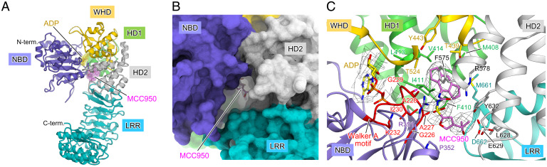
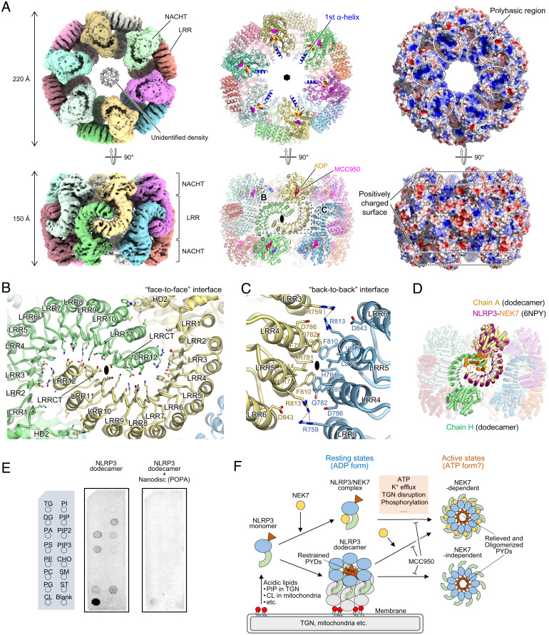
SignificanceThe nucleotide-binding oligomerization domain (NOD)-like receptor pyrin domain containing 3 (NLRP3) is a pattern recognition receptor that forms an inflammasome. The cryo-electron microscopy structure of the dodecameric form of full-length NLRP3 bound to the clinically relevant NLRP3-specific inhibitor MCC950 has established the structural basis for the oligomerization-mediated regulation of NLRP3 inflammasome activation and the mechanism of action of the NLRP3 specific inhibitor. The inactive NLRP3 oligomer represents the NLRP3 resting state, capable of binding to membranes and is likely disrupted for its activation. Visualization of the inhibitor binding mode will enable optimization of the activity of NLRP3 inflammasome inhibitor drugs.

Keywords: NLRP3; NOD-like receptor; inflammasome.


Conflict of interest statement

The authors declare no competing interest.

Figures

Fig. 1.
Fig. 1.
Structure of the human NLRP3ΔP hexamer. (A) Schematic diagram of the domain organization of NLRP3. Residue numbers for human NLRP3 are shown. (B) Size-exclusion chromatography analysis of human NLRP3ΔP and mouse NLRP3 (full length). Elution volumes of the standard proteins thyroglobulin (660 kDa), apoferritin (440 kDa), and bovine serum albumin (134 and 67 kDa) are indicated. (C) Overall structure of the human NLRP3ΔP hexamer. The cryo-EM map (Left) and a ribbon model (Right) are shown. Each protomer is shown in a different color. ADP and MCC950 molecules are shown as yellow and pink spheres, respectively, in the ribbon model. Both threefold and twofold axes are shown. (D and E) Close-up views of the “head-to-tail” (D) and “back-to-back” (E) interfaces. The hydrogen bonds are indicated by dashed lines. The twofold axis is shown in (E). (F) Superposition of the NLRP3-NEK7 complex (PDB 6NPY) onto one protomer in the human NLRP3ΔP hexamer. NLRP3 and NEK7 in the NLRP3-NEK7 complex are shown in dark pink and orange, respectively.
Fig. 2.
Fig. 2.
MCC950 binds the central cavity in the NACHT domain. (A) Protomer structure of the human NLRP3ΔP hexamer. Each domain is shown in different colors according to the color scheme shown in Fig. 1A. ADP and MCC950 molecules are shown as yellow and pink sticks with semitransparent surfaces, respectively. (B) Surface model of the protomer structure in the human NLRP3ΔP hexamer showing the MCC950 binding cavity formed between NBD, HD2, and LRR. (C) Close-up view of the MCC950 binding site. MCC950 and ADP molecules and the side chains of residues surrounding MCC950 are shown in stick models. Hydrogen bond or ionic interactions are indicated by dashed lines. The Walker A motif is highlighted in red.
Fig. 3.
Fig. 3.
Structure of the full-length mouse NLRP3 dodecamer. (A) Overall structure of the full-length mouse NLRP3 dodecamer. Cryo-EM map (Left), ribbon model (Middle), and electrostatic surface potentials (Right) are shown. Each protomer is shown in a different color. ADP and MCC950 molecules are shown as yellow and pink spheres, respectively, in the ribbon model. Both sixfold and twofold axes are shown in the ribbon model. The first α-helix of the NACHT domain is shown in blue in the ribbon model. (B and C) Close-up views of the “face-to-face” (B) and “back-to-back” (C) interfaces. Hydrogen bonds are indicated by dashed lines. The twofold axes are shown. (D) Superposition of the NLRP3-NEK7 complex (PDB 6NPY) onto one protomer in the mouse NLRP3 dodecamer. NLRP3 and NEK7 in the NLRP3-NEK7 complex are shown in dark pink and orange, respectively. (E) Membrane lipid-binding assay of the NLRP3 dodecamer in the absence or presence of POPA-reconstituted nanodiscs (ND). Layout of the membrane lipid strip sheet (Left) and images of the lipid strip sheet detected by chemiluminescence for the NLRP3 dodecamer (Middle) and NLRP3 dodecamer with POPA-reconstituted ND (Right) are shown. TG, triglyceride; DG, diacylglycerol; PA, phosphatidic acid; PS, phosphatidylserine; PE, phosphatidylethanolamine; PC, phosphatidylcholine; PG, phosphatidylglycerol; CL, cardiolipin; PI, phosphatidylinositol; PIP, PtdIns(4)P; PIP2, PtdIns(4,5)P2; PIP3, PtdIns(3,4,5)P3; CHO, cholesterol; SM, sphingomyelin; ST, sulfatide. (F) NLRP3 inflammasome activation model. NEK7-NLRP3 binding and the resting-state oligomerization of NLRP3 (this study) are mutually exclusive events. The NLRP3 dodecamer attaches to the trans-Golgi network (TGN) or mitochondrial membranes via its polybasic region binding to acidic lipids, such as PIP and PA. The NLRP3 inhibitor, MCC950, binds to and stabilizes the closed form of NLRP3, thereby suppressing the disruption of oligomers or structural changes to the open form for NLRP3 activation. Multiple factors are individually or concertedly involved in NLRP3 inflammasome activation, some of which could regulate the activation by directly or indirectly affecting the oligomerized state of NLRP3.

References

    1. Broz P., Dixit V. M., Inflammasomes: Mechanism of assembly, regulation and signalling. Nat. Rev. Immunol. 16, 407–420 (2016). - PubMed
    1. Guo H., Callaway J. B., Ting J. P., Inflammasomes: Mechanism of action, role in disease, and therapeutics. Nat. Med. 21, 677–687 (2015). - PMC - PubMed
    1. Lamkanfi M., Dixit V. M., Mechanisms and functions of inflammasomes. Cell 157, 1013–1022 (2014). - PubMed
    1. Tschopp J., Schroder K., NLRP3 inflammasome activation: The convergence of multiple signalling pathways on ROS production? Nat. Rev. Immunol. 10, 210–215 (2010). - PubMed
    1. Schroder K., Tschopp J., The inflammasomes. Cell 140, 821–832 (2010). - PubMed

MeSH terms

Substances

LinkOut - more resources