. 2022 Mar 7;18(3):e1010354.
doi: 10.1371/journal.ppat.1010354. eCollection 2022 Mar.

β-catenin regulates HIV latency and modulates HIV reactivation

Affiliations

β-catenin regulates HIV latency and modulates HIV reactivation

Hannah J Barbian et al. PLoS Pathog. .

Abstract

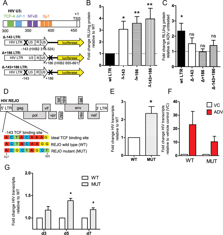
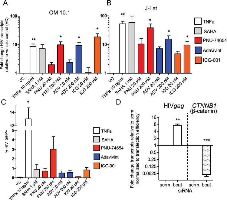
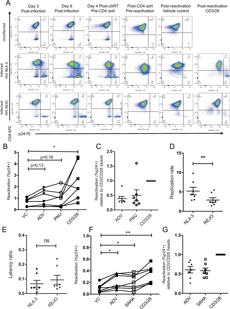
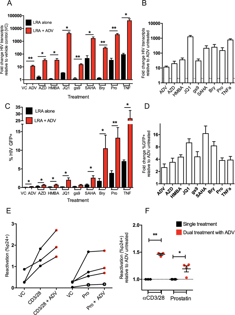
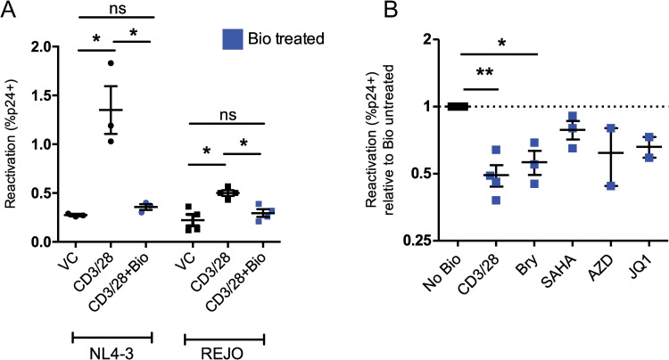
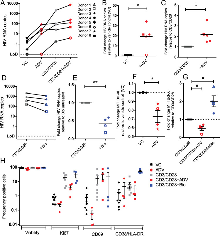
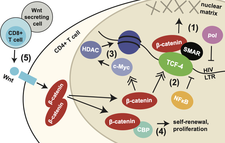
Latency is the main obstacle towards an HIV cure, with cure strategies aiming to either elicit or prevent viral reactivation. While these strategies have shown promise, they have only succeeded in modulating latency in a fraction of the latent HIV reservoir, suggesting that the mechanisms controlling HIV latency are not completely understood, and that comprehensive latency modulation will require targeting of multiple latency maintenance pathways. We show here that the transcriptional co-activator and the central mediator of canonical Wnt signaling, β-catenin, inhibits HIV transcription in CD4+ T cells via TCF-4 LTR binding sites. Further, we show that inhibiting the β-catenin pathway reactivates HIV in a primary TCM cell model of HIV latency, primary cells from cART-controlled HIV donors, and in CD4+ latent cell lines. β-catenin inhibition or activation also enhanced or inhibited the activity of several classes of HIV latency reversing agents, respectively, in these models, with significant synergy of β-catenin and each LRA class tested. In sum, we identify β-catenin as a novel regulator of HIV latency in vitro and ex vivo, adding new therapeutic targets that may be combined for comprehensive HIV latency modulation in HIV cure efforts.


Conflict of interest statement

The authors have declared that no competing interests exist.

Figures

Fig 1
Fig 1. β-catenin modulates HIV transcription in CD4+ T cells via TCF-4 binding at the LTR.
(A) Schematic showing the -143 TCF-4 binding site relative to other transcription factor binding sites on the HIV LTR and with HXB2 numbering, and the LTR luciferase reporter plasmid constructs, containing deleted TCF binding sites shown with X marks at position -143, +186, or both -143 and +186 relative the wildtype HIV BaL transcription start site. (B) CD4+ T cells isolated from n = 5 healthy donors were activated for 24 hrs then nucleofected with LTR reporter constructs. LTR activity was quantified using luciferase relative light units normalized to total protein concentration of the cell lysate. Fold change in LTR activity of the TCF binding site mutants is shown relative to the wildtype HIV BaL LTR control. Nucleofections were performed in duplicate for each donor. (C) CD4+ T cells were nucleofected with LTR reporter constructs as above but were treated with 200 nM adavinint (ADV) 2 hours post-nucleofection. Fold change in LTR activity of wildtype and mutant TCF-4 binding sites are shown relative to cells not treated with ADV. (D) Schematic showing the HXB2 position and sequence of wildtype HIV-REJO TCF binding site compared to the ideal TCF binding site sequence, as well as the mutated TCF binding site in the U3 region of the 5’ LTR of REJO full length molecular clone. (E) CD4+ T cells isolated from n = 6 healthy donors were stimulated for 24 hours then infected with wildtype (WT) or TCF binding site mutant (MUT) HIV REJO via spinoculation with 0.01 MOI virus. Cells were harvested at 24 hours and intracellular HIV transcripts were quantified relative to GAPDH. Fold increase of MUT over WT virus is shown. (F) CD4+ T cells isolated from 4 healthy donors were infected after 24 hours stimulation. 100nM adavivint (ADV) was added following spinoculation, cells were harvested at 24 hours and HIV transcripts quantified as above. Fold change relative of ADV treated cells (red) to DMSO control for wildtype and mutant virus is shown. (G) PBMCs were isolated from 3 healthy donors, stimulated for 3 days, then spinoculated with 0.01 MOI wildtype and TCF binding site mutant virus. Infected cells were harvested at 3, 5, and 7 days post infection, intracellular HIV transcripts were quantified as above. Fold change of TCF binding site mutant to wildtype infected cells harvested on the same day are shown. Columns indicate mean with SEM error bars for all panels. Significance was determined using paired t-tests for all panels, * p<0.05, ** p<0.01.
Fig 2
Fig 2. Inhibition of the β-catenin pathway reactivates HIV in cell lines.
Treatment of OM-10.1 (A) and J-Lat (B) cells with known latency reversing agents TNFα, SAHA, or β-catenin inhibitors PNU-74654 (red), adavivint (blue), and ICG-001 (orange) for 48 hours. Two concentrations of β-catenin inhibitors were tested. Fold change of cellular HIV transcripts relative to DMSO vehicle control is shown, treated conditions were compared to controls from the same experiment. (C) Percent of HIV GFP+ cells following drug treatments are shown. (D) siRNA knockdown of β-catenin in J-Lat cells was performed for 24 hours. Fold change of HIV gag (left) and CTNNB1 (the gene encoding β-catenin, bcat, right) intracellular transcripts to scrambled control siRNA (scrm) is shown (left). Values were normalized to nucleofection efficiency (S1H Fig). Columns indicate mean of 6 (A and B) or 3 (C) replicates with SEM error bars for. Significance was determined using paired t-tests for all panels, * p<0.05, ** p<0.01, *** p<0.001.
Fig 3
Fig 3. β-catenin pathway inhibition reactivates two HIV strains in a primary TCM model of HIV latency.
(A) Surface CD4 and intracellular HIV p24 were measured by flow cytometry throughout the establishment of latently infected TCM cells and following reactivation. Uninfected control (top row), HIV NL4-3 infected (middle row), and HIV REJO infected (bottom row) cultures from one representative donor are shown. (B) Reactivation of latent HIV NL4-3, measured as the percentage of CD4 negative HIV p24 positive cells, in 7 donors treated with β-catenin inhibitors adavivint (ADV, 100 nM), PNU-74654 (100 μM), and positive control αCD3/αCD28 activation beads is shown with lines connecting cells from the same donor. (C) Reactivation of latent NL4-3 with ADV and PNU relative to the % reactivation with αCD3/αCD28 activation beads. (D) The replication rate, measured as the percentage of CD4 negative HIV p24 positive cells at day 6 post-infection over day 3 post-infection, in 7 donors infected with viruses HIV NL4-3 and REJO are shown. (E) The latency ratio, quantified as the percentage of CD4 negative HIV p24 positive cells following reactivation with positive control CD3/28 beads over the percentage of CD4 negative HIV p24 positive cells at peak infection, for 7 donors infected with viruses HIV NL4-3 and REJO are shown. (F) Reactivation of latent HIV REJO, quantified as the percentage of CD4 negative HIV p24 positive cells, for 7 donors treated with β-catenin inhibitor ADV (100 nM) and known latency reversing agents SAHA (1 μM) and CD3/CD28 beads is shown with lines connecting each donor. (G) Reactivation of latent REJO with ADV and SAHA relative to the % reactivation with αCD3/αCD28 activation beads. Statistical p-values were determined via repeated measures ANOVA with Dunnett’s multiple comparisons test for panels B and F, while paired t-test were used for panels c, d, e, and g; * p<0.05, ** p<0.01, *** p<0.001.
Fig 4
Fig 4. β-catenin pathway inhibition enhances HIV latency reversal of other drugs.
(A) Treatment of J-Lat 8.4 cells with established LRAs alone (black) or in combination with 100 nM β-catenin inhibitor ADV (red) for 48 hours. Fold change in HIV gag RNA transcript levels over vehicle control (DMSO) is shown. (B) Fold change in HIV gag RNA levels for LRAs used in combination with ADV, normalized to HIV RNA levels when treated with the LRA alone. (C) Percent of cells positive for intracellular HIV GFP reporter after treatment with LRAs alone (black) or in combination with ADV (red). (D) Fold increase in cells positive for HIV protein (GFP) with ADV treatment, normalized to single LRA treatment. Columns indicate mean of 6 replicates with SEM error bars for (a-d). (E) Latent infection of HIV NL4-3 (closed circles) and REJO (open circles) was modeled in primary CD4+ TCM cells from three to four donors and reactivated with αCD3/αCD28 beads and 1 μM prostatin alone (black) or in combination with 100 nM ADV (red) for 48 hrs. Percent reactivation (percent CD4- p24+ cells) is plotted for each donor with connecting lines. (F) Fold change in the percent of CD4 negative HIV p24 positive cells with combination ADV treatment, normalized to single treatment, is shown. Significance was determined using paired t-tests for all panels, * p<0.05, ** p<0.01.
Fig 5
Fig 5. Activation of β-catenin inhibits HIV latency reversal.
(A) 3–4 human donors were used to establish a primary TCM model of HIV latency using HIV strain NL4-3 (left) or REJO (right). Latently infected cells were stimulated with αCD3/αCD8 activation beads alone (black) or in the presence of 2 μM Bio (blue). The percentage of CD4 negative HIV p24 positive cells is shown. (B) TCM cells latently infected with HIV REJO were stimulated with the indicated drugs in combination with 2 μM Bio (blue). Reactivation as measured by the percentage of CD4 negative HIV p24 positive cells was normalized to reactivation with the drug in the absence of Bio. Mean and SEM of 3 donors is shown. Significance was determined using paired t-tests for all panels, * p<0.05, ** p<0.01.
Fig 6
Fig 6. β-catenin modulation impacts HIV latency reversal in cells from HIV-infected virally suppressed individuals.
(A) CD8-depleted PBMCs from n = 5 HIV positive donors on suppressive cART therapy were treated for 48 hours with 50 nM β-catenin inhibitor ADV, αCD3/αCD28 T-cell activating beads alone or combined with 50 nM ADV. Extracellular (released virions, right) HIV RNA copies were quantified. Absolute RNA copy numbers in vehicle control and ADV treated cultures are shown, with lines connecting samples from the same donor. Symbols corresponding to donors in Table 2 are used for panels a-g. (B) Fold change of RNA copies in ADV treated cultures over vehicle control are shown from released virus. (C) Fold change of RNA copies in ADV co-treated cultures over αCD3/αCD28 single treatment. (D) As in (A), cells were treated with αCD3/αCD28 beads alone or combined with 2 μM 6Bio. HIV RNA quantities are shown with lines connecting cultures from the same donor. (E) Fold change of HIV RNA copies in 6Bio co-treated cells over αCD3/αCD28 single treatment. (f-g) Downstream target of β-catenin, Bcl-xL, was quantified by flow cytometry in cells treated with ADV, αCD3/αCD28, or αCD3/αCD28 with ADV/6Bio, to confirm the modulation of β-catenin by these drugs. Fold change in mean fluorescence intensity of Bcl-xL is shown compared to vehicle control or αCD3/αCD28 treated cells, for single or dual treated cells, respectively. (H) Viability and T cell activation markers in cells following drug treatments were quantified by flow cytometry. The proportion of CD3+ CD4+ T cells expressing Ki67, CD69, CD38/HLA-DR, or LIVE/DEAD stain are plotted for cells treated with the indicated treatments. Significance was determined using paired t-tests for all panels, * p<0.05, ** p<0.01.
Fig 7
Fig 7. Integrated model of potential mechanisms of HIV transcription and latency modulation by β-catenin.
Schematic demonstrating multiple potential mechanisms by which β-catenin may modulate HIV transcription and latency, based on integrated findings from previous studies and data presented here. (1) β-catenin and TCF-4 form a complex with nuclear matrix-associated protein SMAR1 at the HIV LTR just upstream of the transcriptional start site and Sp-1, NFκB, and AP-1 binding sites. This complex pulls the HIV LTR towards the nuclear matrix, occluding access of RNA polymerase [35]. (2) β-catenin positively regulates levels of TCF-4, which has been shown to block binding and transcriptional regulation of NFκB at the HIV LTR [70]. (3) β-catenin further regulates c-Myc levels, which recruit HDAC enzymes, resulting in the viral promoter being more densely packed in chromatin [51]. (4) β-catenin also mediates self-renewal and cell proliferation of memory T cells through CBP, which may contribute to perpetuating the reservoir of latently infected cells [60], (5) A source of β-catenin signaling are CD8+ T cells, which secrete Wnt proteins resulting in stimulation of the Wnt/β-catenin pathway in CD4+ T cells, which culminates in accumulation of β-catenin in the cytoplasm and translocation to the nucleus [33]. This may explain the observed role of CD8+ T cells in maintaining HIV latency. Notably, other cells may serve as a source of Wnt proteins and β-catenin pathway modulating factors.

References

    1. Whitney JB, Hill AL, Sanisetty S, Penaloza-MacMaster P, Liu J, Shetty M, et al.. Rapid seeding of the viral reservoir prior to SIV viraemia in rhesus monkeys. Nature. 2014;512(7512):74–7. doi: 10.1038/nature13594 - DOI - PMC - PubMed
    1. Dufour C, Gantner P, Fromentin R, Chomont N. The multifaceted nature of HIV latency. The Journal of Clinical Investigation. 2020;130(7):3381–90. doi: 10.1172/JCI136227 - DOI - PMC - PubMed
    1. Lorenzo-Redondo R, Fryer HR, Bedford T, Kim E-Y, Archer J, Kosakovsky Pond SL, et al.. Persistent HIV-1 replication maintains the tissue reservoir during therapy. Nature. 2016;530(7588):51–6. doi: 10.1038/nature16933 - DOI - PMC - PubMed
    1. Fletcher CV, Staskus K, Wietgrefe SW, Rothenberger M, Reilly C, Chipman JG, et al.. Persistent HIV-1 replication is associated with lower antiretroviral drug concentrations in lymphatic tissues. Proc Natl Acad Sci U S A. 2014;111(6):2307–12. doi: 10.1073/pnas.1318249111 - DOI - PMC - PubMed
    1. Joseph SB, Kincer LP, Bowman NM, Evans C, Vinikoor MJ, Lippincott CK, et al.. Human Immunodeficiency Virus Type 1 RNA Detected in the Central Nervous System (CNS) After Years of Suppressive Antiretroviral Therapy Can Originate from a Replicating CNS Reservoir or Clonally Expanded Cells. Clinical Infectious Diseases. 2019;69(8):1345–52. doi: 10.1093/cid/ciy1066 - DOI - PMC - PubMed

Publication types