Lifetime changes of the oocyte pool: Contributing factors with a focus on ovulatory inflammation
- PMID: 35255655
- PMCID: PMC8923630
- DOI: 10.5653/cerm.2021.04917
Lifetime changes of the oocyte pool: Contributing factors with a focus on ovulatory inflammation
Abstract
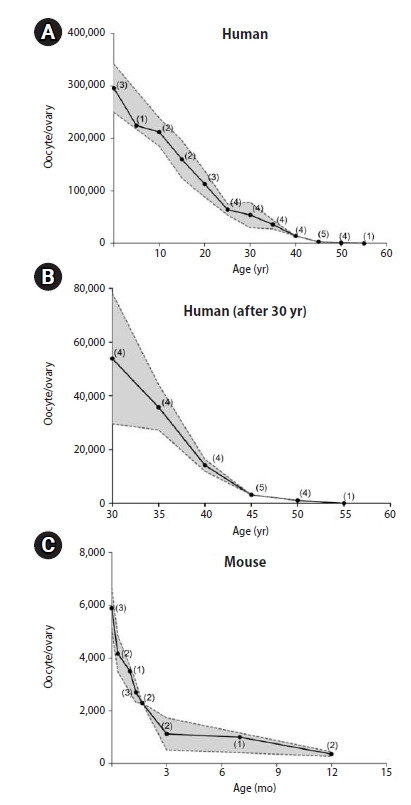
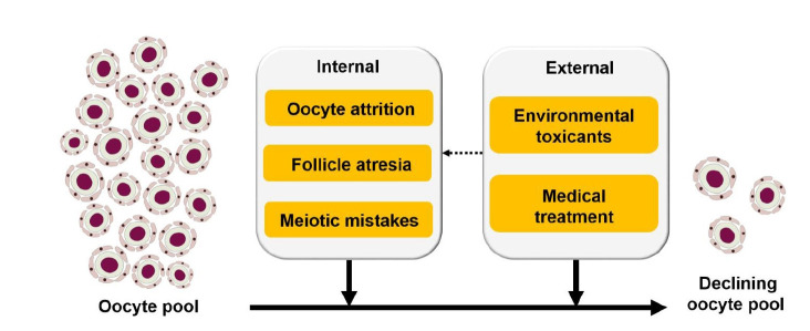
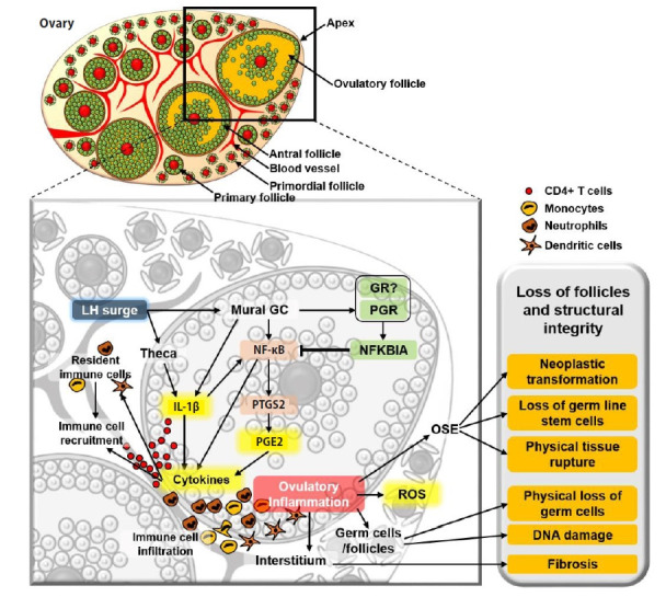
In mammalian species, females are born with a number of oocytes exceeding what they release via ovulation. In humans, an average girl is born with over a thousand times more oocytes than she will ovulate in her lifetime. The reason for having such an excessive number of oocytes in a neonatal female ovary is currently unknown. However, it is well established that the oocyte number decreases throughout the entire lifetime until the ovary loses them all. In this review, data published in the past 80 years were used to assess the current knowledge regarding the changing number of oocytes in humans and mice, as well as the reported factors that contribute to the decline of oocyte numbers. Briefly, a collective estimation indicates that an average girl is born with approximately 600,000 oocytes, which is 2,000 times more than the number of oocytes that she will ovulate in her lifetime. The oocyte number begins to decrease immediately after birth and is reduced to half of the initial number by puberty and almost zero by age 50 years. Multiple factors that are either intrinsic or extrinsic to the ovary contribute to the decline of the oocyte number. The inflammation caused by the ovulatory luteinizing hormone surge is discussed as a potential contributing factor to the decline of the oocyte pool during the reproductive lifespan.
Keywords: Atretic follicle; Folliculogenesis; Germ cells; Inflammation; Oocytes; Reproductive health.
Conflict of interest statement
No potential conflict of interest relevant to this article was reported.
Figures
Similar articles
-
Selective loss of kisspeptin signaling in oocytes causes progressive premature ovulatory failure.Hum Reprod. 2022 Apr 1;37(4):806-821. doi: 10.1093/humrep/deab287. Hum Reprod. 2022. PMID: 35037941 Free PMC article.
-
Postnatal regulation of germ cells by activin: the establishment of the initial follicle pool.Dev Biol. 2006 Oct 1;298(1):132-48. doi: 10.1016/j.ydbio.2006.06.025. Epub 2006 Jun 17. Dev Biol. 2006. PMID: 16930587
-
Pre-ovulatory LH/hCG surge decreases C-type natriuretic peptide secretion by ovarian granulosa cells to promote meiotic resumption of pre-ovulatory oocytes.Hum Reprod. 2011 Nov;26(11):3094-101. doi: 10.1093/humrep/der282. Epub 2011 Aug 23. Hum Reprod. 2011. PMID: 21865234
-
The epidermal growth factor network: role in oocyte growth, maturation and developmental competence.Hum Reprod Update. 2018 Jan 1;24(1):1-14. doi: 10.1093/humupd/dmx029. Hum Reprod Update. 2018. PMID: 29029246 Review.
-
Establishment of oocyte population in the fetal ovary: primordial germ cell proliferation and oocyte programmed cell death.Reprod Biomed Online. 2005 Feb;10(2):182-91. doi: 10.1016/s1472-6483(10)60939-x. Reprod Biomed Online. 2005. PMID: 15823221 Review.
Cited by
-
Environmental Exposures and Polycystic Ovary Syndrome: A Review.Semin Reprod Med. 2024 Dec;42(4):253-273. doi: 10.1055/s-0044-1801405. Epub 2025 Feb 5. Semin Reprod Med. 2024. PMID: 39909399 Review.
-
The Contribution of the Sheep and the Goat Model to the Study of Ovarian Ageing.Biology (Basel). 2023 Feb 8;12(2):270. doi: 10.3390/biology12020270. Biology (Basel). 2023. PMID: 36829547 Free PMC article.
-
Reproductive Ageing: Inflammation, immune cells, and cellular senescence in the aging ovary.Reproduction. 2024 Jun 21;168(2):e230499. doi: 10.1530/REP-23-0499. Print 2024 Aug 1. Reproduction. 2024. PMID: 38744316 Free PMC article. Review.
-
Classification system of human ovarian follicle morphology: recommendations of the National Institute of Child Health and Human Development - sponsored ovarian nomenclature workshop.Fertil Steril. 2025 May;123(5):761-778. doi: 10.1016/j.fertnstert.2024.11.016. Epub 2024 Nov 15. Fertil Steril. 2025. PMID: 39549739
-
Tissue clearing and imaging approaches for in toto analysis of the reproductive system†.Biol Reprod. 2024 Jun 12;110(6):1041-1054. doi: 10.1093/biolre/ioad182. Biol Reprod. 2024. PMID: 38159104 Free PMC article. Review.
References
-
- Block E. Quantitative morphological investigations of the follicular system in women; variations at different ages. Acta Anat (Basel) 1952;14:108–23. - PubMed
-
- Block E. A quantitative morphological investigation of the follicular system in newborn female infants. Acta Anat (Basel) 1953;17:201–6. - PubMed
-
- Faddy MJ, Gosden RG. A mathematical model of follicle dynamics in the human ovary. Hum Reprod. 1995;10:770–5. - PubMed
-
- Forabosco A, Sforza C. Establishment of ovarian reserve: a quantitative morphometric study of the developing human ovary. Fertil Steril. 2007;88:675–83. - PubMed
Grants and funding
LinkOut - more resources
Full Text Sources
Miscellaneous