. 2022 Mar 7;22(1):112.
doi: 10.1186/s12935-022-02466-5.

DNA methyltransferase 1 inhibits microRNA-497 and elevates GPRC5A expression to promote chemotherapy resistance and metastasis in breast cancer

Affiliations

DNA methyltransferase 1 inhibits microRNA-497 and elevates GPRC5A expression to promote chemotherapy resistance and metastasis in breast cancer

Yaobang Liu et al. Cancer Cell Int. .

Abstract

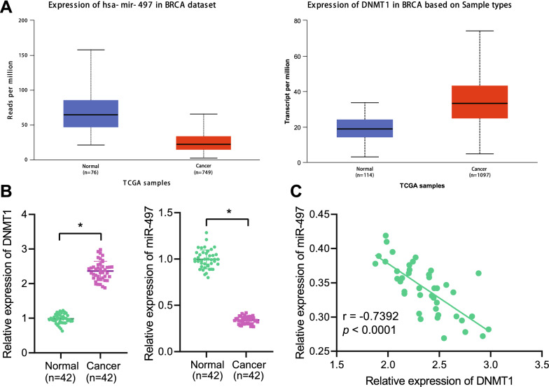
Background: Abnormal DNA methylation of tumor suppressor gene promoter has been found in breast cancer. Therefore, the current study set out to explore how DNA methyltransferase 1 (DNMT1) affects breast cancer through mediating miR-497/GPRC5A axis.

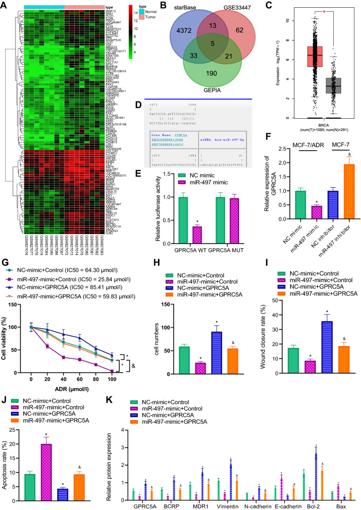
Methods: After loss and gain-of-function approaches were conducted in MCF-7/ADR and MCF-7 cells, cell viability, IC50 value, invasion, migration and apoptosis were measured, respectively. In addition, drug resistance, metastasis and apoptosis-related protein expression were examined using immunoblotting. ChIP and dual-luciferase reporter gene assays were carried out to validate relationship among DNMT1, miR-497, and GPRC5RA. Subcutaneous xenograft tumor model in nude mice was established to detect effects of DNMT1 on growth and metastasis of breast cancer in vivo.

Results: It was found that DNMT1 was notably increased, while miR-497 was poorly-expressed in breast cancer. Highly-expressed DNMT1 could promote chemotherapy resistance and metastasis of breast cancer. Meanwhile, DNMT1 modified methylation of CpG island in miR-497 promoter region, thereby repressing miR-497 level. In addition, miR-497 targeted GPRC5A expression to curb chemotherapy resistance and metastasis of breast cancer cells. Lastly, in vivo experiments showed that knockdown of DNMT1 could suppress breast cancer growth and metastasis.

Conclusions: Collectively, our findings indicated that DNMT1 may inhibit miR-497 and boost the expression of GPRC5A through methylation, thus augmenting breast cancer chemotherapy resistance and metastasis, which provides novel mechanistic insight into the unrecognized roles of DNMT1 in breast cancer.

Keywords: Breast cancer; CpG island; DNA methyltransferase 1; G-protein-coupled receptor family C group 5 member A; MicroRNA-497.


Conflict of interest statement

The authors have no competing interests to declare.

Figures

Fig. 1
Fig. 1
Expression of DNMT1 and miR-497 in breast cancer tissues was examined. A The expression of DNMT1 in 1097 breast cancer samples and 114 adjacent normal tissue samples (left), and expression of miR-497 in 749 breast cancer samples and 76 adjacent normal tissue samples (right) collected in TCGA database. The x-axis represented sample type, the y-axis expression value, and the blue box diagram normal samples. Normal samples were shown in blue box and tumor samples shown in red box. B The expression of DNMT1 and miR-497 in breast cancer tissues and adjacent normal tissues from 42 patients with breast cancer detected by RT-qPCR. C, Pearson correlation analysis adopted to detect the correlation between DNMT1 and miR-497 expression in breast cancer tissues. *p  < 0.05 vs. adjacent normal tissues
Fig. 2
Fig. 2
Effect of DNMT1 on chemotherapy resistance and metastasis of breast cancer cells was examined. A DNMT1 expression in MCF-10A, MCF-7/ADR and MCF-7 cells evaluated using RT-qPCR. B Transduction efficiency in si-DNMT1 or oe-DNMT1-transduced cells determined using RT-qPCR. C The cell viability and IC50 value of ADR detected by CCK-8 assay. D Cell invasion ability detected by Transwell assay. E The migration ability of cells detected by scratch test, and the ratio of scratch area between 48 and 0 h used as the scratch healing rate. F Apoptosis in oe-DNMT1-transduced MCF-7 cells and in si-DNMT1-transduced MCF-7/ADR cells examined using flow cytometry. *p  < 0.05 vs. Oe-NC; &p  < 0.05 vs. si-NC. The experiment was repeated for 3 times independently
Fig. 3
Fig. 3
Regulation of DNMT1 on miR-497 methylation. A The CpG island in the promoter region of miR-497 predicted by MethPrimer website. B The promoter methylation status in 3 cases of breast cancer tissues and adjacent normal tissues detected using MSP. M was the methylation status and U the non-methylation status. C Promoter methylation status in MCF-10A, MCF-7 and MCF-7/ADR cells monitored using MSP. D The methylation status of miR-497 promoter in oe-DNMT1-transduced MCF-10A cells or si-DNMT1-transduced MCF-7 cells examined by MSP. E The expression of miR-497 in oe-DNMT1-transduced MCF-10A cells or si-DNMT1-transduced MCF-7 cells determined using RT-qPCR. F The expression of miR-497 after the treatment of 5-aza-dc detected using RT-qPCR. G The degree of enrichment of DNMT1 on the miR-497 promoter after MCF-7 cells were treated with 5-aza-dc observed by ChIP assay. H The enrichment of H3K27me3 and H3K4me3 in miR-497 promoter region predicted by ENCODE database and validated by ChIP assay in MCF-7 cells. *p  < 0.05 between two groups. The experiment was repeated for 3 times independently
Fig. 4
Fig. 4
DNMT1 regulates the expression of miR-497 through methylation modification and affects breast cancer chemotherapy resistance and metastasis. A The expression of DNMT1 and miR-497 in MCF-7/ADR cells after transduction monitored by RT-qPCR. B The cell viability and IC50 value in MCF-7/ADR cells measured using CCK-8. C Cell invasion of MCF-7/ADR cells detected by means of Transwell assay. D Cell migration of MCF-7/ADR cells examined using scratch assay and the ratio of the scratch area between 48 and 0 h used as the scratch healing rate. E Apoptosis of MCF-7/ADR cells detected using flow cytometry. F Drug resistance, apoptosis and EMT-related protein expression of MCF-7/ADR cells measured by immunoblotting. *p  < 0.05 vs. MCF-7/ADR cells co-transfected with si-NC and NC-inhibitor. &p  < 0.05 vs. MCF-7/ADR cells co-transfected with si-DNMT1 and NC-inhibitor. The experiment was repeated for 3 times independently
Fig. 5
Fig. 5
miR-497 targets and downregulates GPRC5A to affect breast cancer chemotherapy resistance and metastasis. A Heat map of up-regulated gene expression in breast cancer dataset GSE33447. The abscissa represented the sample number, the ordinate the gene name, and the histogram on the upper right was the color scale. B The intersection of miR-497 target genes predicted by StarBase database, markedly up-regulated gene from GSE33447 dataset and up-regulated genes in breast cancer from GEPIA. The middle part represented the intersection of three sets of data. C The differential expression of GPRC5A in breast cancer and normal samples included in TCGA and GTEx. The red box plots in the figure represented tumor samples, and the gray box plots normal samples. D The binding site of miR-497 and GPRC5A predicted using StarBase database. E The binding of miR-497 and GPRC5A verified using dual-luciferase reporter gene assay. *p  < 0.05 vs. NC-mimic group. F The expression of GPRC5A after up-regulation of miR-497 in MCF-7/ADR cells or down-regulation of miR-497 in MCF-7 cells detected using RT-qPCR. *p  < 0.05 vs. NC-mimic-transduced MCF-7/ADR cells. &p  < 0.05 vs. NC-inhibitor-transduced MCF-7/ADR cells. G The viability and IC50 value measured using CCK-8. H Cell invasion detected by means of Transwell assay. I Cell migration examined using scratch assay and the ratio of the scratch area between 48 and 0 h used as the scratch healing rate. J Apoptosis detected using flow cytometry. K Drug resistance, apoptosis and EMT-related protein expression in each group of cells measured by immunoblotting. HK The experiment was repeated for 3 times independently. *p  < 0.05 vs. NC-mimic  +  control. &p  < 0.05 vs. miR-497-mimic  +  control
Fig. 6
Fig. 6
Effects of DNMT1 silencing on the growth and metastasis of breast cancer cells in nude mice. A The efficiency of stable knockdown of DNMT1 in MCF-7/ADR cells detected using RT-qPCR. B Images taken of two groups of nude mice subcutaneously transplanted tumors. C Line graph of tumor volume changes in nude mice, measured once a week. D The weight of tumor tissues in nude mice. E The expression of DNMT1 and miR-497 in tumor tissues of nude mice detected using RT-qPCR. F The expression of related proteins in tumor tissues of nude mice measured using immunohistochemical assay. G Tumor metastasis in nude mice evaluated using in vivo fluorescence imaging. H The dissected lung tissues of nude mice to observe the number of lung metastases. *p  < 0.05 vs. Tissues co-transfected with sh-NC and ADR. n  = 6
Fig. 7
Fig. 7
The molecular mechanism of DNA methyltransferase DNMT1 mediating miR-497/GPRC5A axis and affecting breast cancer chemotherapy resistance and metastasis

References

    1. Dittmer J. Breast cancer stem cells: features, key drivers and treatment options. Semin Cancer Biol. 2018;53:59–74. doi: 10.1016/j.semcancer.2018.07.007. - DOI - PubMed
    1. de Bessa Garcia SA, Araujo M, Pereira T, Mouta J, Freitas R. HOX genes function in breast cancer development. Biochim Biophys Acta Rev Cancer. 2020;1873(2):188358. doi: 10.1016/j.bbcan.2020.188358. - DOI - PubMed
    1. Liang Y, Zhang H, Song X, Yang Q. Metastatic heterogeneity of breast cancer: molecular mechanism and potential therapeutic targets. Semin Cancer Biol. 2020;60:14–27. doi: 10.1016/j.semcancer.2019.08.012. - DOI - PubMed
    1. Lehuede C, Li X, Dauvillier S, Vaysse C, Franchet C, Clement E, Esteve D, Longue M, Chaltiel L, Le Gonidec S, Lazar I, Geneste A, Dumontet C, Valet P, Nieto L, Fallone F, Muller C. Adipocytes promote breast cancer resistance to chemotherapy, a process amplified by obesity: role of the major vault protein (MVP) Breast Cancer Res. 2019;21(1):7. doi: 10.1186/s13058-018-1088-6. - DOI - PMC - PubMed
    1. Petri BJ, Klinge CM. Regulation of breast cancer metastasis signaling by miRNAs. Cancer Metastasis Rev. 2020;39(3):837–886. doi: 10.1007/s10555-020-09905-7. - DOI - PMC - PubMed