

Effect of spermidine on ameliorating spermatogenic disorders in diabetic mice via regulating glycolysis pathway
- PMID: 35255928
- PMCID: PMC8900360
- DOI: 10.1186/s12958-022-00890-w
Effect of spermidine on ameliorating spermatogenic disorders in diabetic mice via regulating glycolysis pathway
Abstract
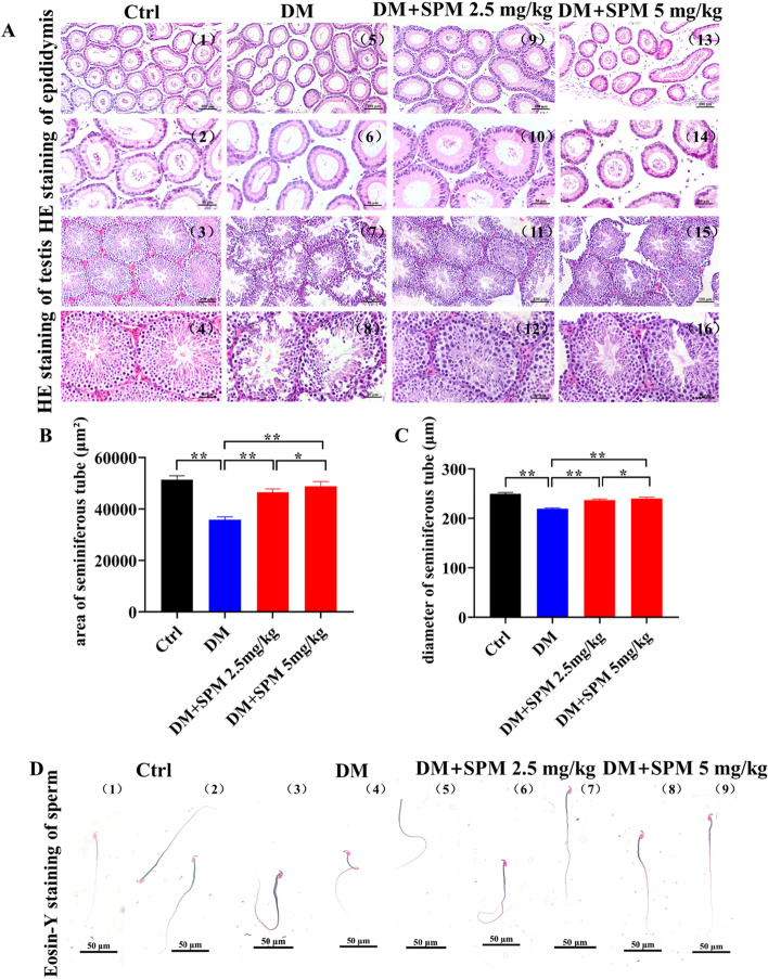
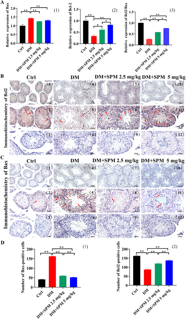
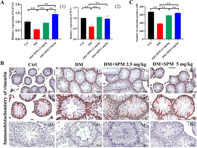
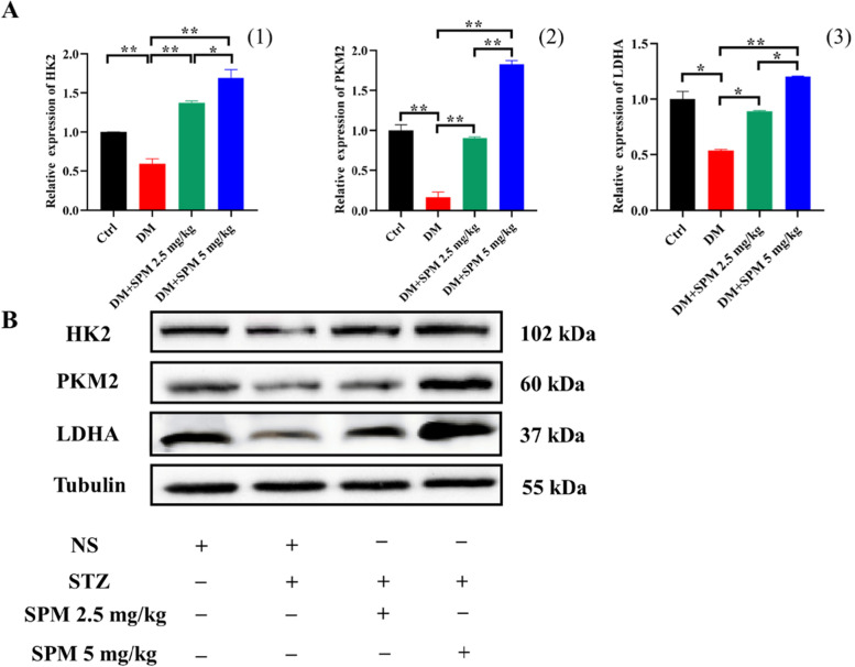
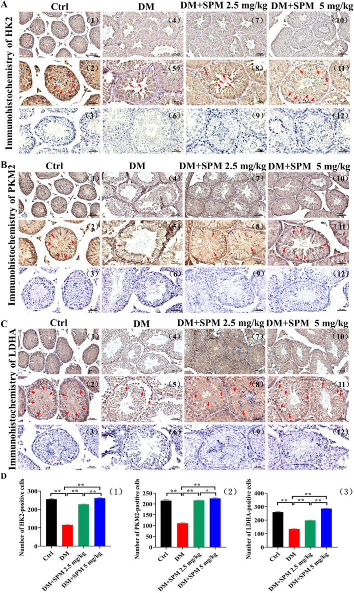
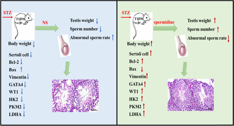
Diabetes mellitus (DM), a high incidence metabolic disease, is related to the impairment of male spermatogenic function. Spermidine (SPM), one of the biogenic amines, was identified from human seminal plasma and believed to have multiple pharmacological functions. However, there exists little evidence that reported SPM's effects on moderating diabetic male spermatogenic function. Thus, the objective of this study was to investigate the SPM's protective effects on testicular spermatogenic function in streptozotocin (STZ)-induced type 1 diabetic mice. Therefore, 40 mature male C57BL/6 J mice were divided into four main groups: the control group (n = 10), the diabetic group (n = 10), the 2.5 mg/kg SPM-treated diabetic group (n = 10) and the 5 mg/kg SPM-treated diabetic group (n = 10), which was given intraperitoneally for 8 weeks. The type 1 diabetic mice model was established by a single intraperitoneal injection of STZ 120 mg/kg. The results showed that, compare to the control group, the body and testis weight, as well the number of sperm were decreased, while the rate of sperm malformation was significantly increased in STZ-induced diabetic mice. Then the testicular morphology was observed, which showed that seminiferous tubule of testis were arranged in mess, the area and diameter of which was decreased, along with downregulated anti-apoptotic factor (Bcl-2) expression, and upregulated pro-apoptotic factor (Bax) expression in the testes. Furthermore, testicular genetic expression levels of Sertoli cells (SCs) markers (WT1, GATA4 and Vimentin) detected that the pathological changes aggravated observably, such as the severity of tubule degeneration increased. Compared to the saline-treated DM mice, SPM treatment markedly improved testicular function, with an increment in the body and testis weight as well as sperm count. Pro-apoptotic factor (Bax) was down-regulated expression with the up-regulated expression of Bcl-2 and suppression of apoptosis in the testes. What's more, expression of WT1, GATA4, Vimentin and the expressions of glycolytic rate-limiting enzyme genes (HK2, PKM2, LDHA) in diabetic testes were also upregulated by SPM supplement. The evidence derived from this study indicated that the SMP's positive effect on moderating spermatogenic disorder in T1DM mice's testis. This positive effect is delivered via promoting spermatogenic cell proliferation and participating in the glycolytic pathway's activation.
Keywords: Diabetes; Glycolytic pathway; Sertoli cells; Spermatogenic dysfunction; Spermidine.
© 2022. The Author(s).
Conflict of interest statement
The authors declare that the research was conducted in the absence of any commercial or financial relationships that could be construed as a potential conflict of interest.
Figures
References
-
- Maresch CC, Stute DC, Alves MG, Oliveira PF, de Kretser DM, Linn T. Diabetes-induced hyperglycemia impairs male reproductive function: a systematic review. Hum Reprod Update. 2018;24:86–105. - PubMed
-
- Xu Y, Wang L, He J, Bi Y, Li M, Wang T, Wang L, Jiang Y, Dai M, Lu J, Xu M, Li Y, Hu N, Li J, Mi S, Chen CS, Li G, Mu Y, Zhao J, Kong L, Chen J, Lai S, Wang W, Zhao W, Ning G, G. China Noncommunicable Disease Surveillance Prevalence and control of diabetes in Chinese adults. JAMA. 2013;310:948–959. - PubMed
-
- Ettinger S. Nutritional pathophysiology of obesity & its comorbidities. 2017. Diabetic nephropathy, chronic kidney disease; pp. 161–189.
-
- Thomas GN, Jiang CQ, Taheri S, Xiao ZH, Tomlinson B, Cheung BM, Lam TH, Barnett AH, Cheng KK. A systematic review of lifestyle modification and glucose intolerance in the prevention of type 2 diabetes. Curr Diabetes Rev. 2010;6:378–387. - PubMed
MeSH terms
Substances
Grants and funding
- Nos. 81860733/The present study was supported by the China National Natural Science Fund
- No. Qian Basic [2019]1344/The Science and Technology Fund of Guizhou Province
- No. 2020JJ5500/Natural Science Foundation Project of Chongqing, Chongqing Science and Technology Commission
- NO. 18B584/Outstanding youth fund of The Education Department of Hunan Province
- No. S202010555129 and X202010555390/Key Laboratory of Engineering Structures Damage and Diagnosis of Hunan Province
LinkOut - more resources
Full Text Sources
Medical
Research Materials
Miscellaneous
