Case Reports
. 2022 Mar 7;15(3):e247900.
doi: 10.1136/bcr-2021-247900.

Breast metastases of eccrine porocarcinoma

Affiliations
Case Reports

Breast metastases of eccrine porocarcinoma

Joana Ferreira Pinto et al. BMJ Case Rep. .

Abstract

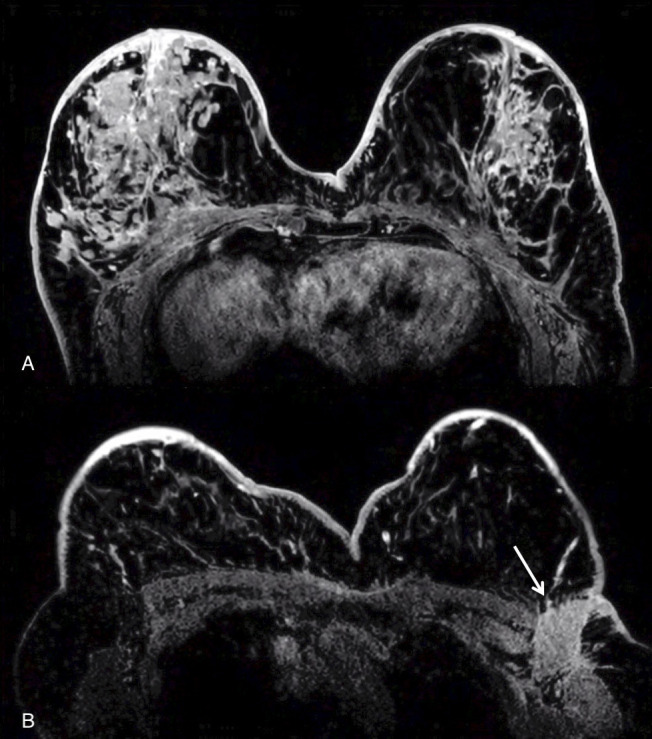
Eccrine porocarcinoma is a rare skin adnexal malignant neoplasm that may arise from a pre-existing benign eccrine poroma or without a predisposing factor. It is a highly invasive neoplasm and has a strong metastatic potential. The most frequently affected organs are the lymph nodes and rarely solid organs such as the liver, lungs and breast. We report a case of a woman with a history of surgically treated eccrine porocarcinoma that a year later presented with multiple lesions in both breasts and axillary lymphadenopathies. After a detailed imaging investigation, the diagnosis of metastatic lesions from porocarcinoma was made. To our knowledge, until the moment, only one case of breast metastasis of eccrine porocarcinoma has been reported in the literature.

Keywords: breast cancer; radiology.


Conflict of interest statement

Competing interests: None declared.

Figures

Figure 1
Figure 1
(A)Axial and (B)sagittal contrast enhanced CT scan showing a 10 cm exophytic epigastric mass with apparent cutaneous origin.
Figure 2
Figure 2
Breast ultrasound (US) demonstrating multiple solid nodular lesions randomly distributed for both breasts (A and B), with greater expression on the right breast and external quadrants of the left breast.
Figure 3
Figure 3
(A) Axial contrast-enhanced T1-weighted images showing multiple bilateral breast lesions with strong enhancement. There is also marked skin thickening, bilateral nipple-areolar-complex invasion and secondary invasion of the pectoral muscle on the right. (B) Axial contrast-enhanced T1-weighted images demonstrating cutaneous involvement by a large left axillary adenopathy.
Figure 4
Figure 4
(A) Low power view of the breast lesion’s core needle biopsy (CNB). Stain: H&E. (B) High-power view of the CNB of the breast lesion: High-grade carcinoma with numerous mitosis and punctuate necrosis. The neoplastic cells have marked nuclear pleomorphism, evident cherry nucleoli, scant cytoplasm with occasional vacuolisation. The histological morphology can be mistaken with high-grade breast carcinoma. (C) Block revision of the skin lesion excision specimen composed of an expansive tumour, partially cystic, with foci of necrosis and epidermal erosion. (D) Higher magnification of the skin lesion excision specimen is composed of a mixed population of clear cells and eosinophilic cells, with obvious atypia and frequent mitotic figures, similar to the morphology of the breast lesion NCB. The similar histology of both lesions, presence of lymphovascular invasion, negative immunohistochemical profile for oestrogen and progesterone receptors, ERBB2, GATA-3, mammoglobin, GCDFP-15, added together to the clinical history were compatible with metastasis to the breast from porocarcinoma of the skin.
Figure 5
Figure 5
(A, E, G) PET-CT showing FDG-avid uptake in both breasts, axillary and cutaneous tumour infiltration and two bone metastases.

References

    1. Mehregan AH, Hashimoto K, Rahbari H. Eccrine adenocarcinoma. A clinicopathologic study of 35 cases. Arch Dermatol 1983;119:104–14. 10.1001/archderm.119.2.104 - DOI - PubMed
    1. Kang M-C, Kim S-A, Lee K-S, et al. . A case of an unusual eccrine poroma on the left forearm area. Ann Dermatol 2011;23:250–3. 10.5021/ad.2011.23.2.250 - DOI - PMC - PubMed
    1. Salih AM, Kakamad FH, Baba HO, et al. . Porocarcinoma; presentation and management, a meta-analysis of 453 cases. Ann Med Surg 2017;20:74–9. 10.1016/j.amsu.2017.06.027 - DOI - PMC - PubMed
    1. Huet P, Dandurand M, Pignodel C, et al. . Metastasizing eccrine porocarcinoma: report of a case and review of the literature. J Am Acad Dermatol 1996;35:860–4. 10.1016/S0190-9622(96)90105-X - DOI - PubMed
    1. Lee SH, Park JM, Kook SH, et al. . Metastatic tumors to the breast: mammographic and ultrasonographic findings. J Ultrasound Med 2000;19:257–62. 10.7863/jum.2000.19.4.257 - DOI - PubMed

Publication types