Review
. 2022 Mar;32(3):344-351.
doi: 10.1136/ijgc-2021-002517.

Updates in the treatment of vaginal cancer

Affiliations
Review

Updates in the treatment of vaginal cancer

Anuja Jhingran. Int J Gynecol Cancer. 2022 Mar.

Abstract

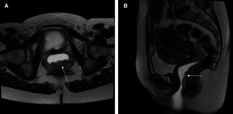
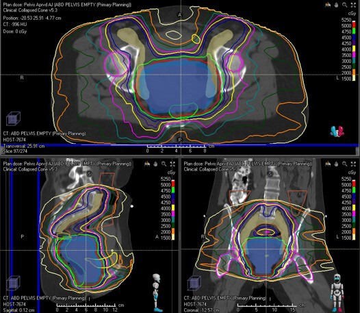
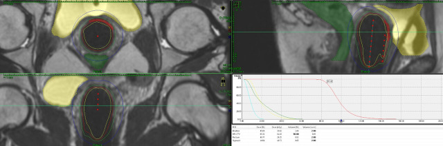
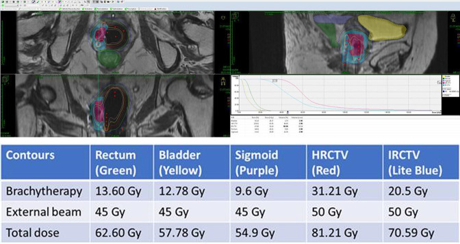
Vaginal cancer is a rare cancer. A lot of the data used in the treatment of this cancer are extrapolated from cervical cancer data. Radiation therapy plays a significant role in the treatment of vaginal cancer. The advances in radiation therapy in both external beam and brachytherapy have improved local control, survival, and toxicity. Brachytherapy plays an important role in treating vaginal cancer, but treatment should be individualized to each tumor. Imaging, particularly magnetic resonance imaging, plays an essential role in the management of patients with vaginal cancer, from diagnosis to staging to treatment management to surveillance.

Keywords: vagina; vaginal fistula; vulvar and vaginal cancer.


Conflict of interest statement

Competing interests: None declared.

Figures

Figure 1
Figure 1
This is a T2 weighted MRI of a patient with vaginal cancer. The lesion shown by the arrow in the posterior wall of the vagina is biopsy-positive vaginal cancer. The lesion involves the entire length of the posterior vaginal wall up to 0.5 cm from the cervix. There is vaginal water base gel in the vagina which shows up as white and separates the vaginal walls so that the lesion can be seen easier.
Figure 2
Figure 2
Volumetric arc therapy for the patient with posterior wall vagina cancer seen in Figure 1. The top image is the axilla view showing the nodal clinical volume (CTV) contours in mustard and the vaginal internal gross tumor volume (GTV) in blue. The nodal CTV which includes the inguinal nodes is receiving 45 Gy and the vaginal GTV is receiving 50 Gy. Normal tissues that are outlined include the bladder in yellow and the rectum in green. The bottom right image is the sagittal view of the same, and bottom left is the coronal view of the same plan.
Figure 3
Figure 3
This figure shows an implant for a patient with a left side vaginal lesion that was <5 mm in depth. A multiple channel cylinder was placed and the needles on the left side as well as the central channel were activated. The high-risk clinical treatment volume received 40.54 Gy from the implant giving it a total of 86.54 Gy with a combination of external beam and brachytherapy. The bladder received 21.79 Gy from the implant for a total of 67.79 Gy, the rectum received 19.37 Gy from the implant for a total of 65.37 Gy, and the sigmoid—which was well away from the implant—received 7.08 Gy from the implant to give it a total of 53.08 Gy.
Figure 4
Figure 4
This figure shows an implant of a patient with right side vaginal tumor. The patient still had residual disease after external beam and therefore a multi-channel cylinder as well as free-hand interstitial needs were used. The needles were placed into the right vaginal wall through the perineum using ultrasound guidance. The planning was done using MRI and CT scan.

Similar articles

Cited by

References

    1. Adhikari P, Vietje P, Mount S. Premalignant and malignant lesions of the vagina. Diagn Histopathol 2017;23:28–34. 10.1016/j.mpdhp.2016.11.006 - DOI
    1. Rajaram S, Maheshwari A, Srivastava A. Staging for vaginal cancer. Best Pract Res Clin Obstet Gynaecol 2015;29:822–32. 10.1016/j.bpobgyn.2015.01.006 - DOI - PubMed
    1. Siegel RL, Miller KD, Jemal A. Cancer statistics, 2020. CA Cancer J Clin 2020;70:7–30. 10.3322/caac.21590 - DOI - PubMed
    1. Serrano B, de Sanjosé S, Tous S, et al. . Human papillomavirus genotype attribution for HPVs 6, 11, 16, 18, 31, 33, 45, 52 and 58 in female anogenital lesions. Eur J Cancer 2015;51:1732–41. 10.1016/j.ejca.2015.06.001 - DOI - PubMed
    1. Sherman JF, Mount SL, Evans MF, et al. . Smoking increases the risk of high-grade vaginal intraepithelial neoplasia in women with oncogenic human papillomavirus. Gynecol Oncol 2008;110:396–401. 10.1016/j.ygyno.2008.05.015 - DOI - PubMed