. 2022 Mar 7;5(6):e202101322.
doi: 10.26508/lsa.202101322. Print 2022 Jun.

Monobodies with potent neutralizing activity against SARS-CoV-2 Delta and other variants of concern

Affiliations

Monobodies with potent neutralizing activity against SARS-CoV-2 Delta and other variants of concern

Taishi Kondo et al. Life Sci Alliance. .

Abstract

Neutralizing antibodies against the severe acute respiratory syndrome coronavirus 2 (SARS-CoV-2) are useful for patients' treatment of the coronavirus disease 2019 (COVID-19). We report here affinity maturation of monobodies against the SARS-CoV-2 spike protein and their neutralizing activity against SARS-CoV-2 B.1.1 (Pango v.3.1.14) as well as four variants of concern. We selected matured monobodies from libraries with multi-site saturation mutagenesis on the recognition loops through in vitro selection. One clone, the C4-AM2 monobody, showed extremely high affinity (K D < 0.01 nM) against the receptor-binding domain of the SARS-CoV-2 B.1.1, even in monomer form. Furthermore, the C4-AM2 monobody efficiently neutralized the SARS-CoV-2 B.1.1 (IC 50 = 46 pM, 0.62 ng/ml), and the Alpha (IC 50 = 77 pM, 1.0 ng/ml), Beta (IC 50 = 0.54 nM, 7.2 ng/ml), Gamma (IC 50 = 0.55 nM, 7.4 ng/ml), and Delta (IC 50 = 0.59 nM, 8.0 ng/ml) variants. The obtained monobodies would be useful as neutralizing proteins against current and potentially hazardous future SARS-CoV-2 variants.


Conflict of interest statement

T Kondo, T Fujino, S Umemoto, G Hayashi, Y Iwatani, and H Murakami are inventors on the provisional patent application (PCT/JP2021/018668; filed 5/18/2021) submitted by Tokai National Higher Education and Research System and National Hospital Organization Nagoya Medical Center. The patent application is for monobody sequences against the SARS-CoV-2 spike protein. Other authors declare that they have no competing interests.

Figures

Figure 1.
Figure 1.. Affinity maturation of the monobodies against SARS-CoV-2 wild type and the neutralizing activity of the matured monobody against the wild type and variants of concern (VOCs).
(A) Development of the matured monobody using TRAP display. Multiple saturation mutagenesis was introduced to the BC and FG loops of the parental monobody, and in vitro selection against the SARS-CoV-2 wild-type receptor-binding domain (RBD) was conducted. The obtained matured monobody had enhanced neutralizing activity against SARS-CoV-2 wild type and VOCs. (B) Structure of the SARS-CoV-2 RBD (PDB entry 6M0J, Lan et al, 2020). The characteristic residues in the VOCs are labeled as K417, L452, T478, E484, and N501. Abbreviations: SARS-CoV-2, severe acute respiratory syndrome coronavirus 2; RBD, receptor-binding domain; ACE2, angiotensin-converting enzyme 2; WT, wild type; VOCs, variants of concern.
Figure 2.
Figure 2.. Affinity maturation of the C4 monobody.
(A) Sequences of the BC and FG loops in the libraries and selected monobodies. Saturation mutagenesis (X) was introduced by NNK codons (N = A, C, G, T; K = G or T; 32 codons/20 aa) at five to six constitutive resides in the BC and FG loops. The probability of amino acids at each position in the loops of the selected clones was shown by WebLogo. The mutated residues in the matured C4-AM1 monobody are highlighted in yellow. The second library was designed to enhance the activity of the C4-AM1 monobody. Partial saturation mutagenesis (X) shown in the WebLogo (gray) was introduced into the library. The mutated residues in the matured C4-AM2 monobody are highlighted in cyan. (B) Selection progress by the TRAP display. A 1 nM receptor-binding domain (RBD) (0.1 pmol) concentration was used for the C4 Lib1-4 selection. The concentration of RBD is stated in the figure used for the C4-AM1 Lib selection. A confirmation experiment (1 nM RBD, 0.5 pmol) was also performed at the 1st, 4th, 7th, and 10th rounds to observe an enrichment of active monobodies. (C) Determination of kinetic parameters by BLI. A biotin-labeled monobody was immobilized on a streptavidin-sensor chip, and SARS-CoV-2 RBD (2.5, 5, 10, 20, and 40 nM) was used in the kinetic analysis. The data are depicted in blue, and the fitted 1:1 binding model in black. The determined kinetic parameters of the monobodies are provided in Table 1. Abbreviations: BLI, Bio-layer interferometry; Lib, library. Other abbreviations are as for Fig 1.
Figure S1.
Figure S1.. Effect of the C4-AM2 monobody’s C-terminal residues.
After purification, the concentration of the C4-AM2 monobodies was adjusted to 150 μM with the buffer B. The buffer was changed to 25 mM Hepes-K, pH 7.5, 150 mM NaCl by gel filtration. UV-vis spectra were measured before (left panel) and after centrifugation (15,300g, 5 min; right panel).
Figure S2.
Figure S2.. Purification of the monobodies.
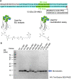
(A) The C4-AM2 monobody sequence. (B) SDS–PAGE analysis of the purified monobodies (150 pmol each) stained using Coomassie Brilliant Blue. After expression in Escherichia coli with 150 ml of Lysogeny Broth and purification using Ni-NTA chromatography, 3–16 mg of the monobody was obtained.
Figure S3.
Figure S3.. Affinity maturation of the C6b monobody.
(A) Sequences of the BC and FG loops in the libraries and selected monobodies. Saturation mutagenesis (X) was introduced by NNK codons (N = A, C, G, T; K = G or T; 32 codons/20 aa) at six to seven constitutive resides in the BC and FG loops. The probability of amino acids at each position in the loops of the selected clones was shown by WebLogo. The mutated residues in the matured C6-AM1 monobody are highlighted in yellow. In addition, other control mutant sequences are shown (PAVT from the original fibronectin, GSGSGS as a linker). The second library was designed to enhance the activity of the C6b monobody. Mutagenesis (X) was introduced by a trinucleotide (19 codons/19 aa). The mutated residues in the matured C4-AM2 monobody are highlighted in cyan. (B) Selection progress by the TRAP display. A 1 nM (0.1 pmol) receptor-binding domain (RBD) concentration was used for the C6b Lib1-3 selection. For the C6b Lib4 selection, a 10 nM (5 pmol) RBD concentration was used for the first round and a 1 nM (0.5 pmol) RBD concentration for subsequent rounds. (C) Determination of kinetic parameters by BLI. A biotin-labeled monobody was immobilized on a streptavidin-sensor chip, and the SARS-CoV-2’s RBD (2.5, 5, 10, 20, and 40 nM) was used in the kinetic analysis. The data are depicted in blue, and the fitting of a 1:1 binding model in black. The determined kinetic parameters of the monobodies are provided in Table 1. *The C6b-AM2 monobody had two amino acids (VR) deletion after the BC loop. Abbreviations are as mentioned in previous figures.
Figure S4.
Figure S4.. Affinity maturation of the C12b monobody.
(A) Sequences of the BC and FG loops in the libraries and selected monobodies. Saturation mutagenesis (X) was introduced by NNK codons (N = A, C, G, T; K = G or T; 32 codons/20 aa) at six to seven constitutive resides in the BC and FG loops. The probability of amino acids at each position in the loops of the selected clones was shown by WebLogo. The mutated residues in the matured C12-AM1 monobody are highlighted in yellow. The second library was designed to enhance the C12b monobody’s activity. Mutagenesis (X) was introduced by a trinucleotide (19 codons/19 aa). The mutated residues in the matured C12-AM2 monobody are highlighted in cyan. (B) Selection progress by the TRAP display. A 1 nM (0.1 pmol) receptor-binding domain (RBD) concentration was used for the C12 Lib1-3 selection. For the C12b Lib4 selection, a 10 nM (5 pmol) RBD concentration was used for the first round, and 1 nM (0.5 pmol) for subsequent rounds. (C) Determination of kinetic parameters by BLI. A biotin-labeled monobody was immobilized on a streptavidin-sensor chip, and SARS-CoV-2 RBD (2.5, 5, 10, 20, and 40 nM) was used in the kinetic analysis. The data are depicted in blue, and the fitting of a 1:1 binding model in black. The determined kinetic parameters of the monobodies are provided in Table 1. * The C12b-AM2 had two amino acids (VR) deletion after the BC loop. Abbreviations are mentioned in the main figures.
Figure 3.
Figure 3.. Binding activity of monobodies against the receptor-binding domains of SARS-CoV-2 wild type and variants of concern analyzed by BLI.
Biotin-labeled monobody was immobilized on a streptavidin-sensor chip, and the receptor-binding domain of SARS-CoV-2 wild type, Alpha, Beta, Gamma, and Delta (2.5, 5, 10, 20, and 40 nM) variants were used in the kinetic analysis. The data are depicted in blue, and the fitted 1:1 binding model in black. The determined kinetic parameters of the monobodies are provided in Tables 1 and 2. The data from Fig 2 (C4, C4-AM2), S3 (C6b), and S4 (C12b) were included to show the complete matrix. Abbreviations are as mentioned previously.
Figure S5.
Figure S5.. Binding activity of the C4-AM2 monobody against the Beta, Gamma, and Delta variants’ receptor-binding domain.
Data in Fig 3 were re-analyzed by a 2:1 heterogeneous ligand model. The determined kinetic parameters of the monobodies are provided in Table S1. Abbreviations are as previously mentioned.
Figure S6.
Figure S6.. Binding activity of the C4 and C4-AM2 monobodies against SARS-CoV-2’s receptor-binding domains carrying a one point mutation observed in the Beta and Delta variants.
A biotin-labeled monobody was immobilized on a streptavidin-sensor chip, and the SARS-CoV-2 (40 nM) mutant receptor-binding domains were used in the kinetic analysis. The data are depicted in blue. Abbreviations are as mentioned in the main figures.
Figure 4.
Figure 4.. Monobody/antibody-mediated neutralization of SARS-CoV-2 wild type and variants of concern infection in VeroE6/TMPRSS cells.
The x-axis value indicates the final concentration of the monobodies (C4, C6b, C12b, and C4-AM2) or the SARS-CoV-2 neutralizing monoclonal antibody, AM-128, for each assay well. The y-axis value displays log10 of each viral RNA copy number in the supernatant. Data are represented as the geometric mean of three independent assays. Abbreviations are as mentioned previously.

Similar articles

Cited by

References

    1. Buyel JF, Twyman RM, Fischer R (2017) Very-large-scale production of antibodies in plants: The biologization of manufacturing. Biotechnol Adv 35: 458–465. 10.1016/j.biotechadv.2017.03.011 - DOI - PubMed
    1. Cao L, Goreshnik I, Coventry B, Case JB, Miller L, Kozodoy L, Chen RE, Carter L, Walls AC, Park YJ, et al. (2020. a) De novo design of picomolar sars-cov-2 miniprotein inhibitors. Science 370: 426–431. 10.1126/science.abd9909 - DOI - PMC - PubMed
    1. Cao Y, Su B, Guo X, Sun W, Deng Y, Bao L, Zhu Q, Zhang X, Zheng Y, Geng C, et al. (2020. b) Potent neutralizing antibodies against sars-cov-2 identified by high-throughput single-cell sequencing of convalescent patients' b cells. Cell 182: 73–84.e16. 10.1016/j.cell.2020.05.025 - DOI - PMC - PubMed
    1. Cathcart AL, Havenar-Daughton C, Lempp FA, Ma D, Schmid MA, Agostini ML, Guarino B, Di iulio J, Rosen LE, Tucker H, et al. . (2021) The dual function monoclonal antibodies VIR-7831 and VIR-7832 demonstrate potent in vitro and in vivo activity against SARS-CoV-2. BioRxiv. 10.1101/2021.03.09.434607. (Preprint posted February 18, 2022) - DOI
    1. Chen P, Nirula A, Heller B, Gottlieb RL, Boscia J, Morris J, Huhn G, Cardona J, Mocherla B, Stosor V, et al. (2021. a) Sars-cov-2 neutralizing antibody ly-cov555 in outpatients with covid-19. N Engl J Med 384: 229–237. 10.1056/NEJMoa2029849 - DOI - PMC - PubMed

Publication types

MeSH terms

Supplementary concepts

Associated data

LinkOut - more resources