. 2022 Mar 7;13(1):1199.
doi: 10.1038/s41467-022-28741-7.

Co-targeting of BAX and BCL-XL proteins broadly overcomes resistance to apoptosis in cancer

Affiliations

Co-targeting of BAX and BCL-XL proteins broadly overcomes resistance to apoptosis in cancer

Andrea Lopez et al. Nat Commun. .

Abstract

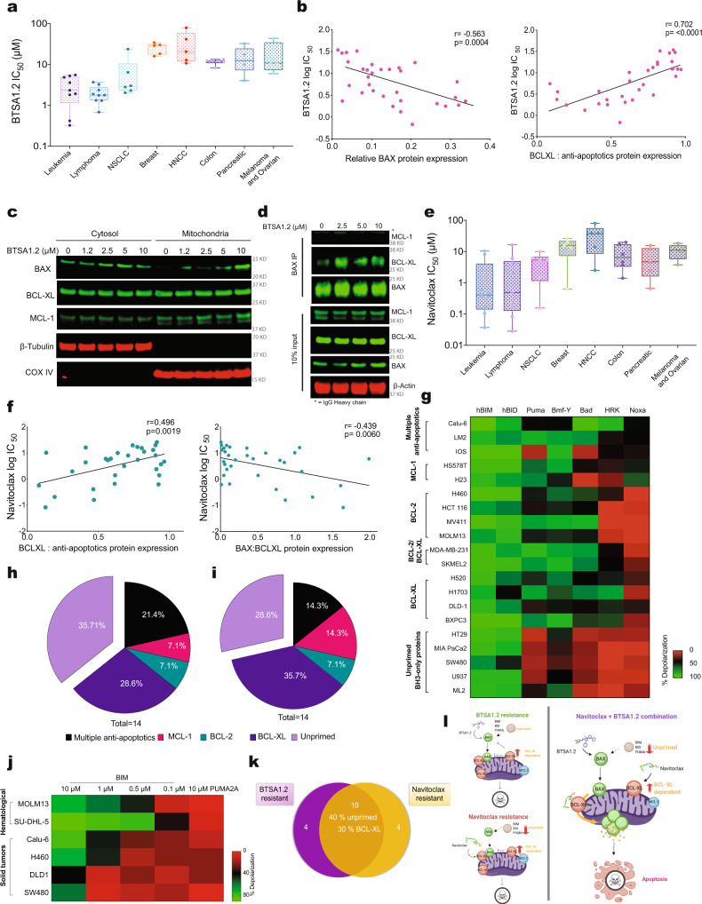
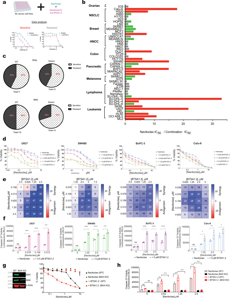
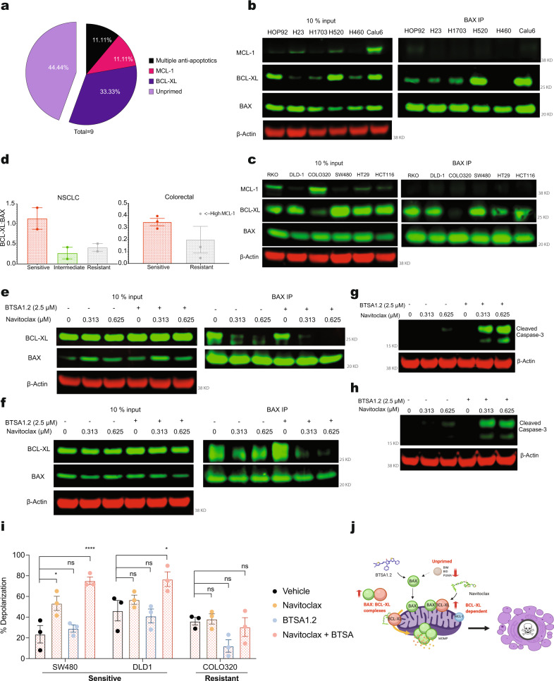
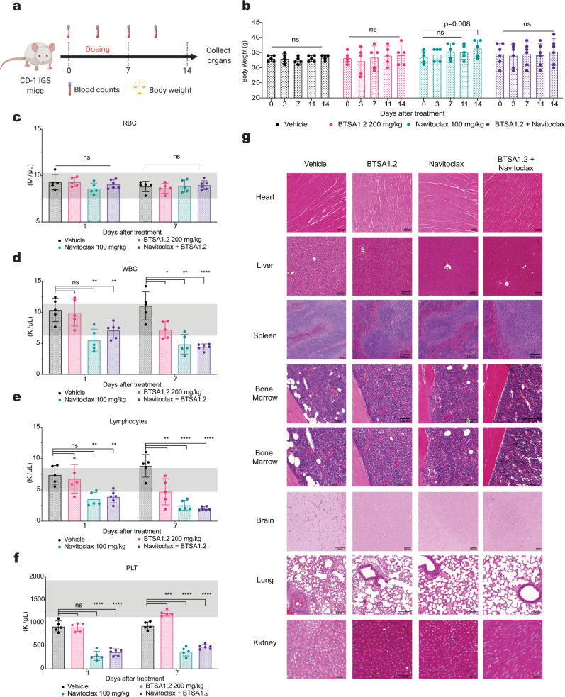
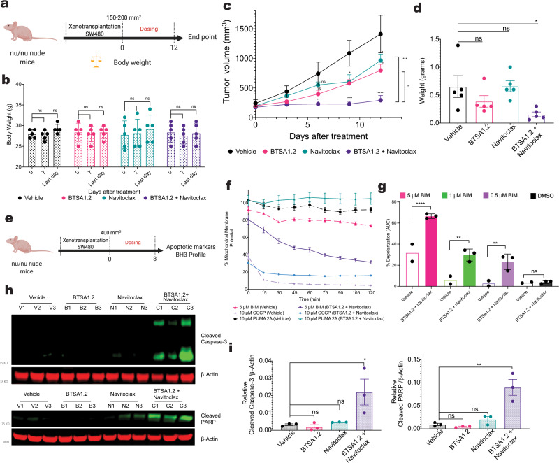
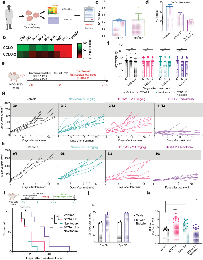
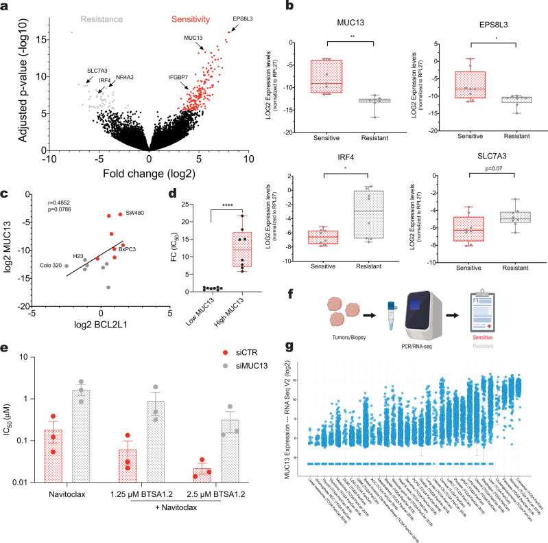
Deregulation of the BCL-2 family interaction network ensures cancer resistance to apoptosis and is a major challenge to current treatments. Cancer cells commonly evade apoptosis through upregulation of the BCL-2 anti-apoptotic proteins; however, more resistant cancers also downregulate or inactivate pro-apoptotic proteins to suppress apoptosis. Here, we find that apoptosis resistance in a diverse panel of solid and hematological malignancies is mediated by both overexpression of BCL-XL and an unprimed apoptotic state, limiting direct and indirect activation mechanisms of pro-apoptotic BAX. Both survival mechanisms can be overcome by the combination of an orally bioavailable BAX activator, BTSA1.2 with Navitoclax. The combination demonstrates synergistic efficacy in apoptosis-resistant cancer cells, xenografts, and patient-derived tumors while sparing healthy tissues. Additionally, functional assays and genomic markers are identified to predict sensitive tumors to the combination treatment. These findings advance the understanding of apoptosis resistance mechanisms and demonstrate a novel therapeutic strategy for cancer treatment.


Conflict of interest statement

E.G., D.E.R., F.K. are inventors on patent applications regarding small molecule BAX activators submitted by Albert Einstein College of Medicine. These patent applications are licensed to BAKX Therapeutics. E.G. is a co-founder and scientific advisor of BAKX Therapeutics, Stelexis Therapeutics, and Selphagy Therapeutics now part of Life Biosciences. E. Vilar has a consulting and advisory role with Janssen Research and Development. None of the above companies have sponsored this research. No potential conflicts of interest were disclosed by the other authors.

Figures

Fig. 1
Fig. 1. Resistance to direct BAX activation and BCL-XL inhibition is regulated by BCL-XL upregulation and an unprimed state.
a A diverse collection of cancer cell lines (n = 46) treated for 72 hrs with BTSA1.2. Box plot corresponds to the tissue type mean cell viability IC50 (μM), cell lines were categorized as sensitive (IC50 < 3 μM) or resistant (IC50 > 3 μM). The lines within the boxes show the median IC50 values, the box denotes the interquartile range (IQR), while the whiskers indicate maxima and minima values. b Correlation of sensitivity to BTSA1.2 with BAX and BCL-XL relative protein levels using Pearson-Correlation. Relative protein levels were normalized to β-Actin loading control, p value was calculated using two-tailed student t-test. c BAX translocation upon 4 h treatment with BTSA1.2 in BxPC-3 cells. d BAX co-IP upon 4 h treatment with BTSA1.2 in BxPC-3 cells. Data are representative of n = 3 independent experiments e, A diverse collection of cancer cells (n = 46) treated for 72 h with Navitoclax. Box plot corresponds to the tissue type mean cell viability IC50 (μM), cell lines were categorized as sensitive (IC50 < 1.5 μM) or resistant (IC50 > 1.5 μM). The lines within the boxes show the median IC50 values, the box denotes the IQR, while the whiskers indicate maxima and minima values f, Correlation of sensitivity to Navitoclax with BCL-XL and BAX:BCL-XL relative protein levels using Pearson-Correlation. Relative protein levels were normalized to β-Actin loading control, p value was calculated using two-tailed student t-test. g Heatmap representation of % mitochondria depolarization of 20 cancer cell lines were classified on different apoptotic blocks based on the BH3 profiling approach. h, i BH3 profiling predicts apoptotic blocks correlated with resistance to h, BTSA1.2 (IC50 > 3 μM) and i, Navitoclax (IC50 > 1.5 μM). j Heatmap representation of mitochondria depolarization upon BIM peptide treatment in hematological and solid tumor malignancies. Puma 2 A peptide was used as negative control. k Venn diagram comparing cell lines resistant to BTSA1.2 and Navitoclax as single agents. l Diagram illustrating the therapeutic strategy of combination treatment with BTSA1.2 and Navitoclax to enhance apoptotic cell death. Data in g an j are mean of three replicates from n = 2 independent experiments. Source data are provided as a Source Data file.
Fig. 2
Fig. 2. BTSA1.2 and Navitoclax synergize to inhibit cell viability and induce apoptosis in various tumor cell lines.
a Schematic of Navitoclax and BTSA1.2 combination screening in a diverse cancer cell lines panel (n = 46). b Bar graph plot of the cell viability IC50 (μM) fold change of cancer cells treated for 72 hrs with Navitoclax in combination with a constant sensitizing concentration of BTSA1.2 (loss of cell viability 0–20%). Red bar graphs correspond to IC50 fold change > 5x; green bar graphs correspond to IC50 fold change 2-4x; and gray bar graphs correspond to IC50 fold change <2x. c Mutation status of TP53 and RAS in cancer cell lines classified as sensitive or resistant to the combination. d Dose-response curves of Navitoclax in the presence of various doses of BTSA1.2 in a panel of cancer cell lines resistant to single agents (Leukemia = U937, Colon=SW480, Pancreatic=BxPC-3, NSCLC = Calu-6). Data are mean ± SD of three technical replicates from n = 3 independent experiments. e Bliss synergy score heat map from combinatorial treatment of BTSA1.2 and Navitoclax in different cancer tissue types in b. Data represent mean from n = 3 independent experiments. f Caspase 3/7 activity assay in diverse cancer cell lines treated with BTSA1.2 and Navitoclax alone or in combination measured at 8 hrs. Data are mean ± SD of three technical replicates from n = 3 independent experiments. Statistics were obtained using two-way ANOVA: *p < 0.05; **p < 0.01; ***p < 0.001; ****p < 0.0001. g Cell viability at 24hrs in WT and CRISPR/Cas9 BAX KO Calu-6 cell lines treated with Navitoclax alone in the presence of a fixed sensitizing concentration of BTSA1.2 (loss of viability <10%). Comparison of BAX and BAK protein expression levels in indicated cell lines. Data are mean ± SD of three technical replicates from n = 3 independent experiments. h Caspase 3/7 activity in WT and CRISPR/Cas9 BAX KO Calu-6 cell lines after 8 hrs treatment with Navitoclax alone and in combination with a fixed sensitizing concentration of BTSA1.2 (loss of viability <10%). Data are mean ± SD of three technical replicates from n = 3 independent experiments. Statistics were obtained using two-way ANOVA: *p < 0.05; **p < 0.01; ***p < 0.001; ****p < 0.0001. Source data are provided as a Source Data file.
Fig. 3
Fig. 3. BAX interaction with BCL-XL dictates sensitivity to BTSA1.2 and Navitoclax combination.
a BH3-profiling predicts apoptotic blocks correlated with sensitivity to the BTSA1.2 and Navitoclax combination. bc Western blot analysis of BAX Co-IP in b, NSCLC and c colorectal cell lines. Representative blot from n = 2 independent experiments. d Quantification of co-immunoprecipitated BAX with BCL-XL in cell lines according to the BTSA1.2 and Navitoclax combination activity (corresponding to Fig. 2a–c). Data is the the normalized values obtained from b, c ± SD from n = 2–3 independent experiments e, f, Western blot analysis of BAX IP in e, NSCLC cell line Calu-6 and f, colorectal cell line SW480 after 4 h treatment with BTSA1.2 and Navitoclax. g, h Detection of cleaved Caspase-3 apoptotic marker by western blot analysis in g, NSCLC cell line Calu6 and h, colorectal cell line SW480 after 4 h treatment with BTSA1.2 and Navitoclax. i Dynamic BH3-profiling of solid tumor cell lines treated with vehicle, Navitoclax, BTSA1.2, or the combination. Bar graph represent % of mitochondria depolarization of cancer cells detected by JC-1 upon treatment with BIM-BH3 derived peptide. Data are mean ± SD of three technical replicates from n = 2 independent experiments. j Schematic of sensitive cells to the BTSA1.2 and Navitoclax combination. Western blot data are a representative of at least three independent experiments. Source data are provided as a Source Data file.
Fig. 4
Fig. 4. Combination of BTSA1.2 and Navitoclax is well tolerated and does not enhance Navitoclax driven toxicity in the hematopoietic system.
a Schematic of BTSA1.2 and Navitoclax combination toxicity study. b Body weight measurements of CD1-IGS mice at 0, 3, 7, 11, and 14 days after the first treatment with vehicle, 100 mg/kg Navitoclax, 200 mg/kg BTSA1.2 or the combination. cf Counts of peripheral c, red blood cells; d white blood cells; e lymphocytes; and f platelets in CD1-IGS mice treated with vehicle, 100 mg/kg Navitoclax, 200 mg/kg BTSA1.2 or the combination at 1 and 7 days after treatment. Normal blood counts range for CD-IGS male mice are indicated in gray. Data in bf, represent mean ± SD from n = 5 mice (Vehicle, BTSA1.2 and Navitoclax) or n = 6 mice (Combination). Statistics were obtained using one-way ANOVA: *p < 0.05; **p < 0.01; ***p < 0.001; ****p < 0.0001. g Representative tissue sections heart, liver, spleen, bone marrow, brain, lung, and kidney using Hematoxylin and Eosin (H&E) staining from mice after treatment of vehicle, 100 mg/kg Navitoclax, 200 mg/kg BTSA1.2 or the combination. Scale bars, 100 μm. Data is representative of n = 3 independent samples per group. Source data are provided as a Source Data file.
Fig. 5
Fig. 5. Combination therapy of BTSA1.2 and Navitoclax shows potent efficacy in resistant colorectal tumor xenografts.
a Schematic of SW480 xenograft efficacy study. b Body weight measurements of Nu/Nu mice at 0, 7, and last day of treatment with vehicle, 100 mg/kg Navitoclax, 200 mg/kg BTSA1.2 or the combination. c Tumor volume curves of vehicle, Navitoclax, BTSA1.2 or the combination cohorts. d Tumor weight after completing study. Data in bd, represent from n = 5 mice (Vehicle, BTSA1.2 and Navitoclax) or n = 6 mice (Combination). Statistics were obtained using two-way Anova: *p < 0.05; **p < 0.01; ***p < 0.001; ****p < 0.0001. e Schematic of SW480 pharmacodynamic xenograft study. f Example of kinetic curve of mitochondria potential in tumors treated with vehicle or combination upon stimuli of BH3-BIM peptide, Puma2A, CCCP or Alamethicin. Data are mean ± SD from n = 3 g, Dynamic BH3-profiling of tumors from mice treated with vehicle or BTSA1.2 and Navitoclax combination. Bar graph represent % of mitochondria depolarization of tumor cells detected by JC-1 upon treatment with BH3-BIM derived peptide or DMSO. Each point corresponds to the mean of n = 3 technical replicates; ± SD from n = 2 independent vehicle mice or n = 3 independent combination mice. Statistics were obtained using two-way Anova: *p < 0.05; **p < 0.01; ***p < 0.001; ****p < 0.0001. h, i Detection of cleaved Caspase-3 and cleaved PARP apoptotic markers by western blot analysis from SW480 tumors. Relative protein levels were normalized to β-Actin loading control. Data are mean ± SD from n = 3 mice. Statistics were obtained using two-way Anova: *p < 0.05; **p < 0.01; ***p < 0.001; ****p < 0.0001. Source data are provided as a Source Data file.
Fig. 6
Fig. 6. Functional markers identify tumors sensitive to the BTSA1.2 and Navitoclax combination.
a Schematic of tumors characterization by BH3-profiling and BAX co-IP to predict efficacy of the BTSA1.2 and Navitoclax combination. b BH3-profiling of colorectal PDXs. Heatmap represent % of mitochondria depolarization of isolated tumor cells detected by JC-1 upon treatment with BH3-derived peptides. Data are mean ± SD from n = 3 independent experiments. c Quantification of co-immunoprecipitated BAX with BCL-XL in colorectal PDX (Supplementary Fig. 13b). Data are mean ± SD from n = 3 independent experiments. d Cell viability of COLO-1 PDX isolated cells after 24 h treatment with 1.25 µM Navitoclax, 10 µM BTSA1.2 or combination. Data are mean ± SD from n = 3 independent experiments. e Schematic of COLO-1 and COLO-2 PDX efficacy study. f Body weight measurements of NOD SCID mice at 0, 6, and last day of treatment with vehicle, 50 mg/kg Navitoclax, 200 mg/kg BTSA1.2 or the combination. Data is the mean ± SD from vehicle n = 9, BTSA1.2, Navitoclax, and combination n = 12 mice body weight. g Tumor volume curves of COLO-1 PDX vehicle, Navitoclax, BTSA1.2 or the combination cohorts. Data in f, g, represents individual measurements (vehicle n = 9, BTSA1.2, Navitoclax, and combination n = 12). h Tumor volume curves of COLO-2 PDX vehicle, Navitoclax, BTSA1.2 or the combination cohorts. Data represents individual measurements (vehicle n = 5, BTSA1.2, Navitoclax, and combination n = 8). i Survival of COLO-1 PDX after 18 days of treatment with vehicle, 50 mg/kg Navitoclax, 200 mg/kg BTSA1.2 or the combination, n = 8. j Dynamic BH3-profiling of COLO-1 tumors from mice treated with vehicle or BTSA1.2 and Navitoclax combination. Bar graph represent % of mitochondria depolarization of tumor cells detected by JC-1 upon treatment with BH3-BIM, BH3-BID or Puma2A derived peptide. Each data point is the mean of n = 3 technical replicates per samples; ± SD from n = 2 independent mice tumor samples. k Relative BCL-XL levels of COLO-1 tumors. Relative protein levels were normalized to β-Actin loading control. Data are mean ± SD from n = 8 mice. Statistics were obtained using one-way Anova: *p < 0.05; **p < 0.01; ***p < 0.001; ****p < 0.0001. Source data are provided as a Source Data file.
Fig. 7
Fig. 7. Bioinformatic analysis predicts markers of sensitivity and resistance to the BTSA1.2 and Navitoclax combination.
a Volcano plot showing the expression change and significance level of genes between sensitive and resistant cell lines as defined based on the IC50 change from Navitoclax alone to BTSA1.2 and Navitoclax combination (corresponding to Fig. 2b). Top 250 predicted markers of sensitivity (red) and resistance (gray) are highlighted. b Box plot of validation of top hits associated with sensitivity and resistance to the BTSA1.2 and Navitoclax combination (corresponding to Fig. 2b) by RT-qPCR. Relative gene expression was normalized using RPL27. Analysis done in 7 sensitive cell lines and 6 resistant cell lines. The lines within the boxes show the median expression values, the box denotes the IQR, while the whiskers indicate maxima and minima values. Data are mean ± SD from n = 3 independent experiments. Statistics were obtained using two-tailed student t-test: *p < 0.05; **p < 0.01; ***p < 0.001; ****p < 0.0001. c Correlation of BCL2L1 (refers to BCL-XL protein) relative gene expression levels and MUC13 gene expression levels in cell lines categorized as sensitive or resistant to the BTSA1.2 and Navitoclax combination (corresponding to Fig. 2b) using Pearson-Correlation. d Box plot of MUC13 expression grouped by the sensitivity to the BTSA1.2 and Navitoclax combination (corresponding to Fig. 2b). The lines within the boxes show the median MUC13 expression values, the box denotes the IQR, while the whiskers indicate maxima and minima values. Data are mean ± SD from n = 8 independent experiments. Statistics were obtained using two-tailed student t-test: *p < 0.05; **p < 0.01; ***p < 0.001; ****p < 0.0001. e Bar graph plot of the cell viability IC50 (μM) of SW480 after transfection with siRNA CTR or siRNA MUC13 and treated for 24 h with Navitoclax in combination with a constant sensitizing concentration of BTSA1.2 (loss of cell viability <20%). Data are mean ± SD from n = 3 independent experiments. f Schematic of various tumors analyzed for gene expression to predict sensitivity to the BTSA1.2 and Navitoclax combination. g MUC13 cancer patient’s expression data using TCGA and other non-redundant data from cbioportal.org. Statistics were obtained using student t-test: *p < 0.05; **p < 0.01; ***p < 0.001; ****p < 0.0001. Source data are provided as a Source Data file.

Similar articles

Cited by

References

    1. Hanahan D, Weinberg RA. Hallmarks of cancer: the next generation. Cell. 2011;144:646–674. - PubMed
    1. Delbridge ARD, Grabow S, Strasser A, Vaux DL. Thirty years of BCL-2: translating cell death discoveries into novel cancer therapies. Nat. Rev. Cancer. 2016;16:99–109. - PubMed
    1. Singh R, Letai A, Sarosiek K. Regulation of apoptosis in health and disease: the balancing act of BCL-2 family proteins. Nat. Rev. Mol. Cell Biol. 2019;20:175–193. - PMC - PubMed
    1. Hata AN, Engelman JA, Faber AC. The BCL2 family: key mediators of the apoptotic response to targeted anticancer therapeutics. Cancer Discov. 2015;5:475–487. - PMC - PubMed
    1. Llambi F, Green DR. Apoptosis and oncogenesis: give and take in the BCL-2 family. Curr. Opin. Genet. Dev. 2011;21:12–20. - PMC - PubMed

Publication types

MeSH terms