

Ultrasound-guided needle positioning for nodal dynamic contrast-enhanced MR lymphangiography
- PMID: 35256625
- PMCID: PMC8901837
- DOI: 10.1038/s41598-022-07359-1
Ultrasound-guided needle positioning for nodal dynamic contrast-enhanced MR lymphangiography
Abstract
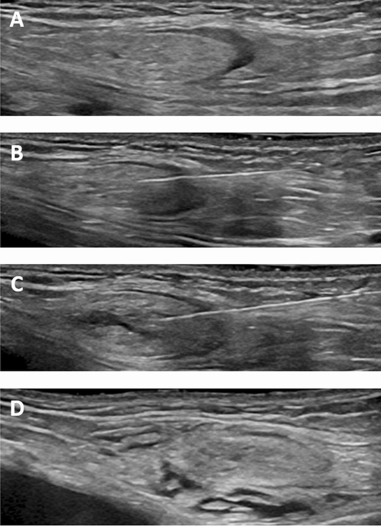
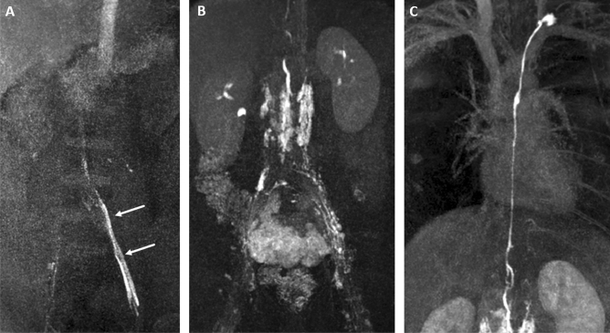
The aim of the study was to assess injection needle positioning for contrast-enhanced MR-lymphangiography (MRL) by ultrasound-guided injection of saline-solution. 80 patients (33 male, mean age 43.1 years) were referred for MRL. The injection needle position was assessed by injection of saline-solution. Consecutive lymph node distension was observed on sonography followed by MRL. Transpedal MRL was performed when no inguinal lymph nodes could be identified. The inguinal lymph node detection rate was recorded. MR-lymphangiograms were assessed regarding primary (i.e. enhancement of draining lymph vessels) and secondary technical success (i.e. lymph vessel enhancement after repositioning of the needle). MRL was considered as clinically successful if enhancement of the central lymphatic system and/or a lymphatic pathologies were observed. For a total of 92 MRLs 177 groins were evaluated sonographically. In 171/177 groins (96.6%) lymph nodes were identified. After needle placement lymph node distension was observed in 171/171 cases (100%) on saline injection. MR-contrast injection demonstrated enhancement of draining lymph vessels in 163/171 cases (95.3%). In 6/171 cases (3.5%) in-bore needle retraction lead to lymphatic enhancement. In one patient [2/171 nodes (1.1%)] no lymphatic enhancement was seen despite repeated needle repositioning. Overall contrast application was technically successful in 169/171 cases (98.8%). In the 6 groins in which no nodes were identifiable, transpedal MRL was successful. So overall 91/92 MRLs (98.9%) were clinically successful. No complications were recorded. Confirmation of the needle position for nodal MRL by sonographically controlled saline injection is a reliable technique with a high success rate of MRL.
© 2022. The Author(s).
Conflict of interest statement
The authors declare no competing interests.
Figures
References
MeSH terms
Substances
LinkOut - more resources
Full Text Sources
