. 2022 Mar 7;12(1):3687.
doi: 10.1038/s41598-022-07642-1.

UCHL5 controls β-catenin destruction complex function through Axin1 regulation

Affiliations

UCHL5 controls β-catenin destruction complex function through Axin1 regulation

Wonhee Han et al. Sci Rep. .

Abstract

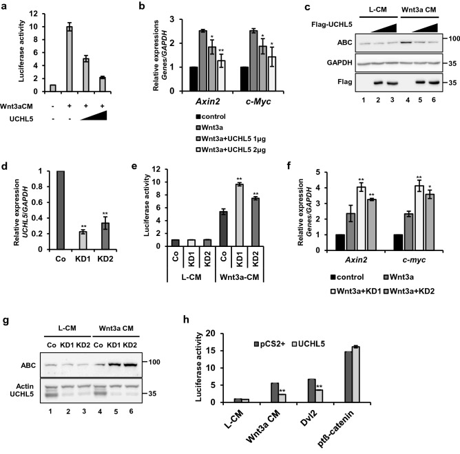
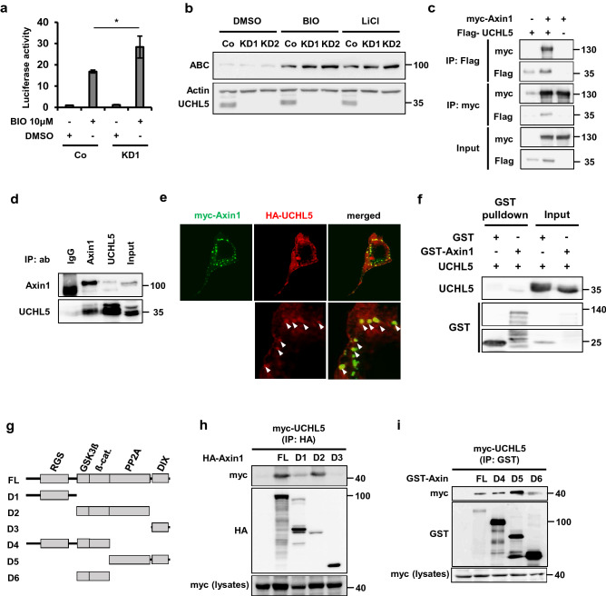
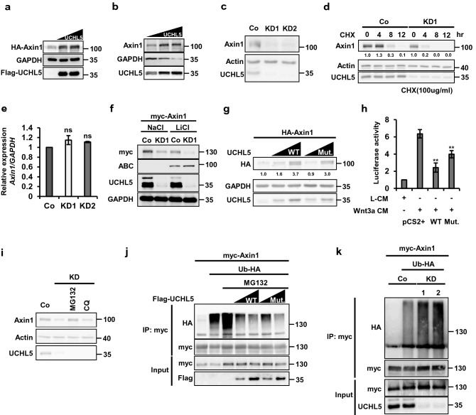
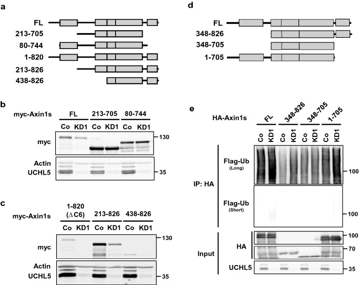
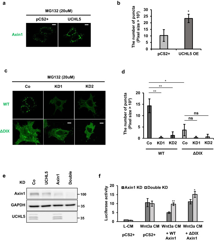
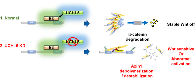
Wnt/β-catenin signaling is crucially involved in many biological processes, from embryogenesis to cancer development. Hence, the complete understanding of its molecular mechanism has been the biggest challenge in the Wnt research field. Here, we identified ubiquitin C-terminal hydrolase like 5 (UCHL5), a deubiquitinating enzyme, as a novel negative regulator of Wnt signaling, upstream of β-catenin. The study further revealed that UCHL5 plays an important role in the β-catenin destruction complex, as it physically interacts with multiple domains of Axin1 protein. Our functional analyses also elucidated that UCHL5 is required for both the stabilization and the polymerization of Axin1 proteins. Interestingly, although these events are governed by deubiquitination in the DIX domain of Axin1 protein, they do not require the deubiquitinating activity of UCHL5. The study proposes a novel molecular mechanism of UCHL5 potentiating the functional activity of Axin1, a scaffolder of the β-catenin destruction complex.


Conflict of interest statement

The authors declare no competing interests.

Figures

Figure 1
Figure 1
UCHL5 negatively regulates the Wnt/ß-catenin signaling pathway. (a) TOPflash assay using HeLa cells. Cells were transfected with the indicated plasmids (50 ng TK-Renilla reporter; 250 ng TOPFlash; 250 ng FOPFlash; 1 μg and 2 μg pCS2 + UCHL5). Then, Wn3a conditioned media (Wnt3a CM) was treated for 16 h. (b) Quantitative PCR (qPCR) analysis for Axin2, pCS2 + UCHL5 was introduced into HeLa and MCF7 cells. Then, Wnt3a CM was treated for 16 h. GAPDH was used for normalization. (c) Western blot analysis using HeLa cells. Flag-UCHL5 plasmids (2 μg and 4 μg) were transfected with empty vector. Transfected cells were treated with either L- cell control CM (L CM) or Wnt3a CM for 16 h. (d) qPCR analysis for UCHL5. Total RNAs were extracted from control (Co) HeLa and two different stable knockdown HeLa cells (KD1 and KD2). Samples were subjected to cDNA synthesis and qPCR analysis. GAPDH was used for normalization. (e) TOPflash assay using stable HeLa cells. Co, KD1, and KD2 HeLa cells were transfected with TOPflash (200 ng) and TK-Renilla reporter (50 ng). Then, cells were treated with Wnt3a CM for 16 h. (f) qPCR analysis for Axin2 and c-myc. Co, KD1, and KD2 HeLa cells were incubated with Wnt3a CM for 16 h. (g) Western blot analysis using Co and KD HeLa cells. Co, KD1 and KD2 HeLa cells were incubated with Wnt3a CM for 16 h. (h) TOPflash assay using Co and KD1 HeLa cells. Either pCS2 + or pCS2 + UCHL5 (1 μg) was co-transfected with TOPflash reporter (250 ng) and TK-Renilla reporter (50 ng) into cells. For Wnt stimulation, Wnt3a CM (for 16 h), Dvl plasmids (0.5 μg), and ptß-catenin plasmids (0.2 μg) were introduced. The data from TOPflash (a, e, h) and qPCR analyses (b, d, f) are displayed as means ± SD and show a representative of multiple independent experiments (n = 3 biological independent experiments). * P < 0.05, ** P < 0.005. from two-tailed unpaired t-test (a, b,d, e, f, h).
Figure 2
Figure 2
UCHL5 interacts with Axin1. (a) TOPflash assay using Co and KD1 HeLa cells. Co and KD1 HeLa cells were transfected with TOPflash (200 ng) and TK-Renilla reporter (50 ng). Then, cells were treated with BIO (1 μM) for 16 h. The data is displayed as means ± SD and show a representative of multiple independent experiments (n = 3 biological independent experiments). * P < 0.05. (b) Western blot analysis using Co and KD HeLa cells. BIO (2 μM) and LiCl (25 Mm) were added to Co, KD1 and KD2 HeLa cell culture media for 6 h. (c) Co-IP assay using HeLa cells. Indicated plasmids were introduced to cells (2 μg myc-Axin1; 2 μg Flag-UCHL5). The transfected cells were treated with MG132 (5 μM, 16 h) and then subjected to immunoprecipitation using anti-Flag and anti-myc. (d) Co-IP assay using HeLa cells. After treatment with MG132 (5 μM, 16 h), cells were lysed and precipitated with the indicated antibodies (anti-IgG, anti-Axin1, and anti-UCHL5). (e) Immunocytochemistry assay. HeLa cells were transfected with myc-Axin1 (200 ng) and HA-UCHL5 plasmids (200 ng). Transfected cells were then fixed and stained with anti-myc (green) and anti-HA (red). Scale bar represents 10 μm. (f). (b) In vitro binding assay using recombinant proteins. UCHL5 and GST-Axin1 proteins were incubated and then subjected to immunoblotting. (g) Diagram of a series of Axin1 deletion mutants used for IP assay. (h, i) IP assay using HeLa cells. Cells were transfected with Flag-UCHL5 (2 μg) and various truncated Axin1 mutants (2 μg D1-D6).
Figure 3
Figure 3
UCHL5 stabilizes Axin1 in a non-enzymatic fashion. (a) Western blot analysis using HeLa cells. Cells were transfected with HA-Axin1 (0.5 μg) and Flag-UCHL5 plasmids (2 μg and 3 μg). Cell lysates were then immunoblotted to detect the level of HA-Axin1, Flag-UCHL5, and GAPDH proteins. (b) Western blot analysis using HeLa cells. Flag-UCHL5 plasmids (2 μg and 3 μg) were introduced into cells. Cell lysates were then subjected to immunoblotting to detect endogenous Axin1, Flag-UCHL5, and GAPDH proteins. (c) Western blot analysis using Co, KD1, and KD2 HeLa cells. Endogenous Axin1, Actin, and UCHL5 levels were determined by anti-Axin1, anti-Actin, and anti-UCHL5. (d) Pulse-chase test using HeLa cells. The level of endogenous Axin1 protein was measured over time under the translation-blocked condition by cycloheximide (100 μg/ml). The band intensity of Axin1 was measured with ImageJ software and was normalized to Actin. (e) qPCR analysis for the expression of Axin1 mRNA in Co, KD1, and KD2 HeLa cells. The level of Axin1 mRNA was normalized by GAPDH mRNA. (f) Western blot analysis using Co and KD1 HeLa cells. Cells were transfected with myc-Axin1 (1 μg), and treated with either NaCl or LiCl (50 mM). Cell lysates were then subjected to immunoblotting to detect myc-Axin1, active ß-catenin, UCHL5, and GAPDH proteins. (g) Western blot analysis using HeLa cells. The requirement of deubiquitinating activity for the Axin1 stabilization was determined by comparing WT and Mut.UCHL5. HA-Axin1 (0.5 μg), WT UCHL5 (1 μg and 2 μg), and Mut. UCHL5 (1 μg and 2 μg) were introduced into KD1 HeLa cells. Cell lysates were immunoblotted with antibodies against HA, Actin, and UCHL5. The band intensity of HA-Axin1 was measured with ImageJ software and was normalized to GAPDH. (h) TOPflash assay using HeLa cells. Cells were transfected with TOPflash reporter (250 ng) and TK-Renilla reporter (50 ng), together with either WT UCHL5 or Mut.UCHL5. 8 h after transfection, cell culture media were replaced with Wnt3a CM and cells were further incubated for 16 h. (i) Western blot analysis using HeLa cells. KD1 HeLa cells were treated with either MG132 (20 μM) and Chloroquine (100 μM) for 5 h. Then, cell lysates were immunoblotted with antibodies for Axin1, UCHL5, and Actin. (j) In vivo ubiquitination assay using HeLa cells. myc-Axin1 (2 μg) was transfected alone or with Ub-HA (2 μg), together with either Flag-UCHL5 (2 μg and 4 μg) or Flag-Mut. UCHL5 (2 μg and 4 μg). After that, Cells were treated with MG132 (20 μM) for 5 h and then lysed and subjected to immunoprecipitation with anti-myc. (k) In vivo ubiquitination assay using HeLa cells. myc-Axin1 (2 μg) was transfected into WT and two KD HeLa cells (KD1 and KD2), alone or with Ub-HA (2 μg). Transfected cells were treated with MG132 (20 μM) for 5 h and then subjected to immunoprecipitation with anti-myc. The data from qPCR analysis (e) and TOPflash (h) and displayed as means ± SD and show a representative of multiple independent experiments (n = 3 biological independent experiments). ns, not significant and ** P < 0.005. from two-tailed unpaired t-test (e, h).
Figure 4
Figure 4
DIX domain is crucial for UCHL5-mediated Axin1 stabilization. (a) Diagram of a series of Axin1 deletion mutants used for western blotting. (b,c) 1 μg of myc- full-length Axin1 (FL) and truncated Axin1 mutants (213–705 a.a., 80–744 a.a., 1–820 a.a., 213–826 a.a., 438–826 a.a.) were transfected into WT and KD1 HeLa cells. Resulting cells lysates were subjected to immunoblotting with antibodies against myc, Axin, and UCHL5. (d) Diagram of a series of Axin1 deletion mutants used for ubiquitination assay. (e) In vivo ubiquitination assay using Co and KD1 HeLa cells. HA-Axin1 (2 μg) and Ub-Flag (2 μg) were transfected into WT and two KD1 HeLa cells. The transfected cells were treated with MG132 (20 μM) for 5 h and then subjected to immunoprecipitation with anti-HA.
Figure 5
Figure 5
UCHL5 is required for DIX-mediated polymerization of Axin1. (a) Immunocytochemistry assay. GFP-Axin1 plasmids (400 ng) were introduced into cells with either empty vector (2 μg) or UCHL5 plasmids (2 μg). The transfected cells were treated with MG132 (5 μM) for 16 h. Then, the cells were fixed and immunostained with anti-GFP and Alexa 488 mouse secondary antibody. Scale bars represent 10 μm. (b) Quantification of the GFP puncta average number from the images in (a) performed using ImageJ quantification tool. Quantification performed from 3 experiments with 5 cells quantified for each condition. Data are displayed as means ± SD and show a representative of three independent experiments. *P < 0.05 from two-tailed t-test. (c) Immunocytochemistry assay. GFP-Axin1 plasmids (400 ng) were introduced into WT and KD cells (KD1 and KD2). Then, the transfected cells were treated with MG132 (5 μM) for 16 h. After that, the cells were fixed and immunostained as described in a. Scale bars represent 10 μm. (d) Quantification of the GFP puncta average number from the images in (c) performed using ImageJ quantification tool. Quantification performed from 3 experiments with 5 cells quantified for each condition. Data are displayed as means ± SD and show a representative of three independent experiments. ns, not significant. *P < 0.05, **P < 0.005, from two-tailed t-test. (e) Western blot analysis using HeLa cells. Lysates from Axin1-deficient (Axin1 KD) and Axin1/UCHL5-double deficient cells (Double KD) were immunoblotted with anti-Axin1, anti-UCHL5, and anti-GAPDH. (f) TOPflash assay using HeLa cells. Either Axin1 KD or Double KD HeLa cells were transfected with TOPflash reporter (250 ng) and TK-Renilla reporter (50 ng), together with either wild-type Axin1 (500 ng) or DIX-deficient Axin1 (ΔDIX; 500 ng). 8 h after transfection, cell culture media were changed with Wnt3a CM and cells were further incubated for 16 h. The data is displayed as means ± SD and show a representative of multiple independent experiments (n = 3 biological independent experiments). *P < 0.05, **P < 0.005.
Figure 6
Figure 6
UCHL5 is required for the functional activity of Axin1. UCHL5 blocks ubiquitination of Axin1 DIX domain. As a result, Axin1 is stabilized and successfully polymerized. However, absence of UCHL5 promotes DIX ubiquitination and results in failure of Axin1 polymerization and stabilization. The image was generated using Microsoft PowerPoint software Version 1907 (Build 11901.20176).

References

    1. Routledge D, Scholpp S. Mechanisms of intercellular Wnt transport. Development. 2019 doi: 10.1242/dev.176073. - DOI - PubMed
    1. Jiang X, Cong F. Novel regulation of Wnt signaling at the proximal membrane level. Trends Biochem. Sci. 2016;41:773–783. doi: 10.1016/j.tibs.2016.06.003. - DOI - PubMed
    1. Schaefer KN, Peifer M. Wnt/beta-catenin signaling regulation and a role for biomolecular condensates. Dev. Cell. 2019;48:429–444. doi: 10.1016/j.devcel.2019.01.025. - DOI - PMC - PubMed
    1. Anthony CC, Robbins DJ, Ahmed Y, Lee E. Nuclear Regulation of Wnt/beta-catenin signaling: It's a complex situation. Genes (Basel) 2020 doi: 10.3390/genes11080886. - DOI - PMC - PubMed
    1. Kim SE, et al. Wnt stabilization of beta-catenin reveals principles for morphogen receptor-scaffold assemblies. Science. 2013;340:867–870. doi: 10.1126/science.1232389. - DOI - PMC - PubMed

Publication types