. 2022 Mar 7;12(1):3622.
doi: 10.1038/s41598-022-07603-8.

Bordetella pertussis-infected innate immune cells drive the anti-pertussis response of human airway epithelium

Affiliations

Bordetella pertussis-infected innate immune cells drive the anti-pertussis response of human airway epithelium

M M Kroes et al. Sci Rep. .

Abstract

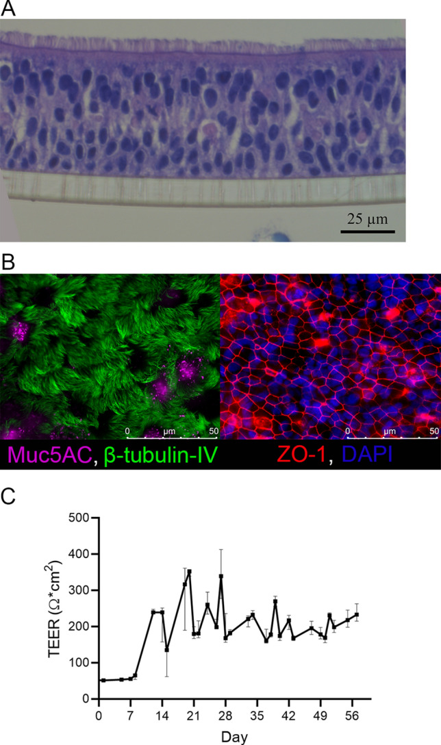
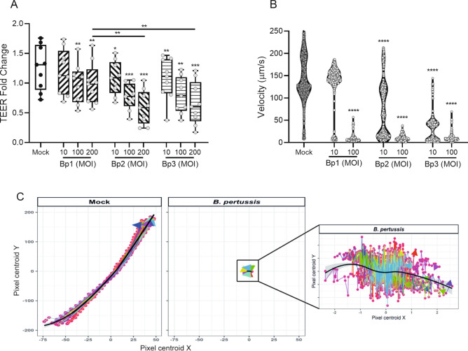
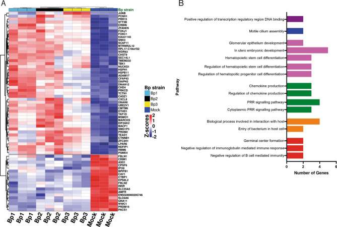
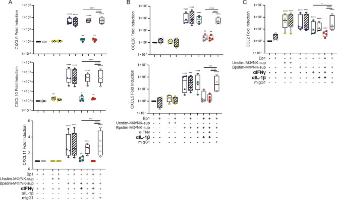
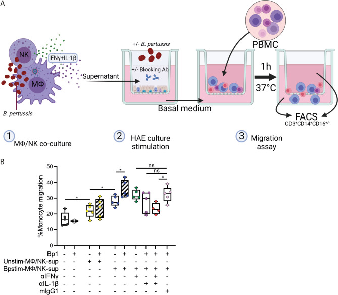
Pertussis is a severe respiratory tract infection caused by Bordetella pertussis. This bacterium infects the ciliated epithelium of the human airways. We investigated the epithelial cell response to B. pertussis infection in primary human airway epithelium (HAE) differentiated at air-liquid interface. Infection of the HAE cells mimicked several hallmarks of B. pertussis infection such as reduced epithelial barrier integrity and abrogation of mucociliary transport. Our data suggests mild immunological activation of HAE by B. pertussis indicated by secretion of IL-6 and CXCL8 and the enrichment of genes involved in bacterial recognition and innate immune processes. We identified IL-1β and IFNγ, present in conditioned media derived from B. pertussis-infected macrophage and NK cells, as essential immunological factors for inducing robust chemokine secretion by HAE in response to B. pertussis. In transwell migration assays, the chemokine-containing supernatants derived from this HAE induced monocyte migration. Our data suggests that the airway epithelium on its own has a limited immunological response to B. pertussis and that for a broad immune response communication with local innate immune cells is necessary. This highlights the importance of intercellular communication in the defense against B. pertussis infection and may assist in the rational design of improved pertussis vaccines.


Conflict of interest statement

The authors declare no competing interests.

Figures

Figure 1
Figure 1
HAE cultures. (A) HE staining of HAE cultures differentiated for 7 weeks. (B) Fluorescence microscopy images of HAE cultures differentiated for 7 weeks and stained with (left panel) β-tubulin IV (Cilia, green) and Muc5AC (Mucus, pink) or (right panel) ZO-1 (Tight junction, red) and DAPI (Nuclei, blue). (C) TEER development during HAE culturing in time (days). Data are represented as median and 95% confidence interval of 4 different donors from 4 independent experiments.
Figure 2
Figure 2
Barrier function and mucociliary clearance after stimulation with three different B. pertussis strains. (A) TEER of HAE cultures after 22 h in medium only (Mock, black dots) or with B. pertussis stimulation (grey dots) relative to the TEER prior stimulation. Data are represented as boxplots and the filling represents different B. pertussis strains used and individual dots are averages of triplicate measurements. The data shown are from 4 donors measured twice across 3 independent experiments. (B) Velocity of beads moving over the apical surface of HAE cells after a 22 h Mock (black) or B. pertussis (grey) stimulation. Data are represented as violin plots, dots represent individual bead trajectories. The data shown are from 2 donors from 3 independent experiments. Significance represented relative to Mock-treated HAE cells. *p < 0.05, **p < 0.01, ***p < 0.001, ****p < 0.0001. (C) Normalized directional bead displacement of Mock- (left panel) or B. pertussis- (middle and right panel) stimulated HAE cells. Individual dots represent individually detected beads and arrows indicate trajectory of beads, black line indicate average bead locations. Data shown are of one representative location in one Mock- or B. pertussis-stimulated HAE cells.
Figure 3
Figure 3
Transcriptome analysis of B. pertussis-infected HAE cells. (A) Heatmap representing hierarchical clustering of all up- or downregulated genes (padj < 0.05, rows; z-scores, scaled and centered per gene) after a 6 h stimulation with any of the three used B. pertussis strains per individual HAE donor (columns; Mock (dark blue), Bp1 (light blue), Bp2 (black) and Bp3 (yellow)). (B) GO enrichment analysis of B. pertussis-infected HAE cells representing number of upregulated genes present (x-axis) in enriched pathways (y-axis). Padj = 0.119 for all enriched pathways. Data are shown from transcriptome analysis from 3 individual donors.
Figure 4
Figure 4
IL-6 and CXCL8 secretion by HAE cells stimulated with three different B. pertussis strains. (A) IL-6 and (B) CXCL8 secretion in the basal medium of HAE cultures after 22 h of stimulation. Data are represented as fold change of cytokine levels relative to that of the Mock-treated HAE cells. Box filling (stripes diagonal left, diagonal right and horizontal) represents the different B. pertussis strains used and individual dots are averages of triplicate measurements. The data shown are from 3 donors measured twice across 3 independent experiments. Significance represented relative to Mock-treated HAE cells. *p < 0.05, **p < 0.01, ***p < 0.001, ****p < 0.0001.
Figure 5
Figure 5
Cytokine-dependent chemokine secretion by HAE cells. (A) IFNγ-dependent chemokine secretion (CXCL9, CXCL10, CXCL11), (B) IL-1β-dependent chemokine secretion (CCL5, CCL20, CXCL5, CXCL8) and (C) IFNγ or IL-1β-dependent chemokine secretion (CCL2) by HAE cells after 22 h of stimulation in the absence of cytokines (black, grey) or in the presence of 10 ng/ml IFNγ (green), IL-1β (purple), TNF (orange) or all three cytokines (red) in the absence (open boxplot) or presence (diagonally striped boxplot) of Bp1 MOI 100. Data are represented as fold change of cytokine levels relative to that of the Mock-treated HAE cells. Individual dots are averages of triplicate measurements. The data shown are from 3 donors measured twice across 3 independent experiments. Significance represented relative to Mock-treated HAE cells, unless indicated otherwise. *p < 0.05, **p < 0.01, ****p < 0.0001.
Figure 6
Figure 6
Chemokine secretion by HAE cells in response to stimulation with supernatants derived from B. pertussis- or Mock-treated macrophage/NK co-cultures. (A) IFNγ-dependent chemokine secretion (CXCL9, CXCL10, CXCL11), (B) IL-1β-dependent chemokine secretion (CCL20, CXCL5) and (C) IFNγ, IL-1β or TNF-dependent chemokine secretion (CCL2) by HAE cells after 22 h of stimulation in the absence of MΦ/NK-sup (black, grey) or in the presence of Unstim-MΦ/NK-sup (yellow) or Bpstim-MΦ/NK-sup in the absence of blocking antibodies (blue) or in the presence of αIFNγ (green), αIL-1β (purple), both (red), or the isotype control mIgG1 (pink). All these conditions were performed in the absence (open boxplot) or presence (diagonally striped boxplot) of Bp1 MOI 100. Data are represented as fold change of chemokine levels relative to that of the Mock-treated HAE cells. Individual dots are averages of triplicate measurements. The data shown are from 3 donors measured twice across 2 independent experiments. Unstim-MΦ/NK-sup = supernatant derived from unstimulated macrophage/NK cell co-cultures, Bpstim-MΦ/NK-sup = supernatant derived from B. pertussis-infected macrophage/NK cell co-cultures. Significance represented relative to Mock-treated HAE cells, unless indicated otherwise. *p < 0.05, **p < 0.01, ***p < 0.001, ****p < 0.0001.
Figure 7
Figure 7
Monocyte migration in response to stimulated HAE culture basal medium. (A) Graphical representation of the PBMC migration experiment. Image created with BioRender.com (B) % of monocyte migration in response to the basal medium derived from HAE cultures after 22 h of stimulation in the absence of MΦ/NK-sup (black, grey) or in the presence of Unstim-MΦ/NK-sup (yellow) or Bpstim-MΦ/NK-sup in the absence of blocking antibodies (blue) or in the presence of αIFNγ (green), αIL-1β (purple), both (red), or the isotype control mIgG1 (pink). All these conditions were performed in the absence (open boxplot) or presence (diagonally striped boxplot) of Bp1 MOI 100. Data are represented as the amount of CD3-CD14+CD16+/- cells in the basal compartment relative to the total amount of CD3-CD14+CD16+/- cell in the basal and apical compartment. Individual dots are averages of triplicate measurements. Representative data shown of HAE basal medium derived from 4 donors from 4 independent experiments used in 2 independent PBMC migration assays. *p < 0.05.
Figure 8
Figure 8
Graphical representation of the human airway epithelium response to B. pertussis. Upon infection of the HAE, B. pertussis disrupts the epithelial barrier integrity and mucociliary clearance (1). Local innate immune cells such as macrophages and NK cells sense the presence of B. pertussis and produce cytokines including, IFNγ and IL-1β (2). These cytokines stimulate the HAE to produce a wide variety of chemokines (3) which are required for the recruitment of immune cells to the site of infection (4) amplifying local inflammation. Image created with BioRender.com.

References

    1. Paddock CD, et al. Pathology and pathogenesis of fatal bordetella pertussis infection in infants. Clin. Infect. Dis. 2008;47:328–338. doi: 10.1086/589753. - DOI - PubMed
    1. Yeung KHT, Duclos P, Nelson EAS, Hutubessy RCW. An update of the global burden of pertussis in children younger than 5 years: A modelling study. Lancet Infect. Dis. 2017;17:974–980. doi: 10.1016/s1473-3099(17)30390-0. - DOI - PubMed
    1. van der Lee S, Hendrikx LH, Sanders EAM, Berbers GAM, Buisman AM. Whole-cell or acellular pertussis primary immunizations in infancy determines adolescent cellular immune profiles. Front. Immunol. 2018;9:51. doi: 10.3389/fimmu.2018.00051. - DOI - PMC - PubMed
    1. Ryan M, et al. Distinct T-cell subtypes induced with whole cell and acellular pertussis vaccines in children. Immunology. 1998;93:1–10. doi: 10.1046/j.1365-2567.1998.00401.x. - DOI - PMC - PubMed
    1. Hovingh ES, et al. Emerging Bordetella pertussis strains induce enhanced signaling of human pattern recognition receptors TLR2, NOD2 and secretion of IL-10 by dendritic cells. PLoS ONE. 2017;12:e0170027. doi: 10.1371/journal.pone.0170027. - DOI - PMC - PubMed

Publication types