Review
. 2022 Feb;12(2):511-531.
doi: 10.1016/j.apsb.2021.06.014. Epub 2021 Jun 30.

Targeting whole body metabolism and mitochondrial bioenergetics in the drug development for Alzheimer's disease

Affiliations
Review

Targeting whole body metabolism and mitochondrial bioenergetics in the drug development for Alzheimer's disease

Steven N Austad et al. Acta Pharm Sin B. 2022 Feb.

Abstract

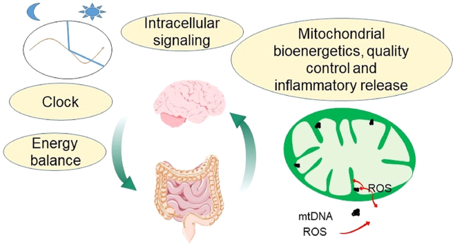
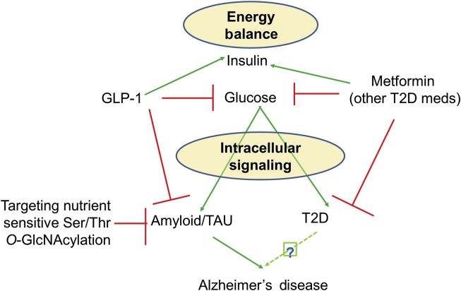
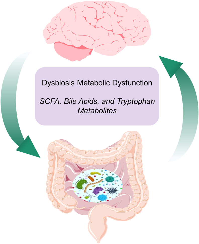
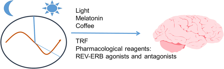
Aging is by far the most prominent risk factor for Alzheimer's disease (AD), and both aging and AD are associated with apparent metabolic alterations. As developing effective therapeutic interventions to treat AD is clearly in urgent need, the impact of modulating whole-body and intracellular metabolism in preclinical models and in human patients, on disease pathogenesis, have been explored. There is also an increasing awareness of differential risk and potential targeting strategies related to biological sex, microbiome, and circadian regulation. As a major part of intracellular metabolism, mitochondrial bioenergetics, mitochondrial quality-control mechanisms, and mitochondria-linked inflammatory responses have been considered for AD therapeutic interventions. This review summarizes and highlights these efforts.

Keywords: ACE2, angiotensin I converting enzyme (peptidyl-dipeptidase A) 2; AD, Alzheimer's disease; ADP, adenosine diphosphate; ADRD, AD-related dementias; Aβ, amyloid β; CSF, cerebrospinal fluid; Circadian regulation; DAMPs; DAMPs, damage-associated molecular patterns; Diabetes; ER, estrogen receptor; ETC, electron transport chain; FCCP, trifluoromethoxy carbonylcyanide phenylhydrazone; FPR-1, formyl peptide receptor 1; GIP, glucose-dependent insulinotropic polypeptide; GLP-1, glucagon-like peptide-1; HBP, hexoamine biosynthesis pathway; HTRA, high temperature requirement A; Hexokinase biosynthesis pathway; I3A, indole-3-carboxaldehyde; IRF-3, interferon regulatory factor 3; LC3, microtubule associated protein light chain 3; LPS, lipopolysaccharide; LRR, leucine-rich repeat; MAVS, mitochondrial anti-viral signaling; MCI, mild cognitive impairment; MRI, magnetic resonance imaging; MRS, magnetic resonance spectroscopy; Mdivi-1, mitochondrial division inhibitor 1; Microbiome; Mitochondrial DNA; Mitochondrial electron transport chain; Mitochondrial quality control; NLRP3, leucine-rich repeat (LRR)-containing protein (NLR)-like receptor family pyrin domain containing 3; NOD, nucleotide-binding oligomerization domain; NeuN, neuronal nuclear protein; PET, fluorodeoxyglucose (FDG)-positron emission tomography; PKA, protein kinase A; POLβ, the base-excision repair enzyme DNA polymerase β; ROS, reactive oxygen species; Reactive species; SAMP8, senescence-accelerated mice; SCFAs, short-chain fatty acids; SIRT3, NAD-dependent deacetylase sirtuin-3; STING, stimulator of interferon genes; STZ, streptozotocin; SkQ1, plastoquinonyldecyltriphenylphosphonium; T2D, type 2 diabetes; TCA, Tricarboxylic acid; TLR9, toll-like receptor 9; TMAO, trimethylamine N-oxide; TP, tricyclic pyrone; TRF, time-restricted feeding; cAMP, cyclic adenosine monophosphate; cGAS, cyclic GMP/AMP synthase; hAPP, human amyloid precursor protein; hPREP, human presequence protease; i.p., intraperitoneal; mTOR, mechanistic target of rapamycin; mtDNA, mitochondrial DNA; αkG, alpha-ketoglutarate.


Figures

Image 1
Graphical abstract
Figure 1
Figure 1
Relationships among energy balance, insulin/glucose, intracellular insulin signaling, targeting protein Ser/Thr O-GlcNAcylation, type two diabetes (T2D), and AD risk. Green arrows indicate “stimulation” and red lines “inhibition”.
Figure 2
Figure 2
A common link between metabolic dysregulation and AD is dysbiosis of the human intestinal tract or “gut”—commonly defined as a disturbance of or change in the density and/or composition of gut microbiota. Bacterial metabolites (purple box), may influence metabolism to directly or indirectly impact neuronal function. Overall, the gut–brain axis (the brain affects the gut through neuronal and hormonal signals, and the gut microbiome affects the brain) is an important integrative system that modulates metabolic balance and, hence, is a potential target for managing AD.
Figure 3
Figure 3
The circadian clock regulates metabolism and mitochondrial function in various model systems. Disturbances in the sleep/wake cycle in aging people and AD patients were prominent disruptive symptoms. Thus, restricted light, restricted food intake, and reagents that modulate the circadian clock, such as melatonin, coffee, and REV-ERB agonists and antagonists, are being considered for targeting whole-body and mitochondrial metabolism in the context of AD therapy.
Figure 4
Figure 4
Currently, more women than men have AD. However, this may be due to socioeconomic or lifespan differences between men and women. It has been shown that men with AD die earlier than women with AD. Even though there is no difference in lifespan between sexes, mitochondrial complex I and II activities are different between sexes in the 3xTg AD model. In the hAPP model, the X chromosome has been shown to extend survival and enhance cognition.
Figure 5
Figure 5
Compounds or peptides that target mitochondrial complex I, complex V, mitochondrial fission protein DRP1; and means to deliver mitochondrial protease, and TCA cycle substrates have been tested in preserving/improving mitochondrial function or in AD models.
Figure 6
Figure 6
Examples of compounds that enhances mitophagy. Urolithin A and UMI-77 have been shown to improve cognition and decrease pathology in the APP/PS1 model of AD. Urolithin A increases mitophagy by increasing PINK1 and Parkin and improves bioenergetics in cells and worms. UMI-77 enhances MCL-1 and LC3 interaction on mitochondria, and show benefits in attenuating cognitive deficits and pathology in APP/PS1 mice. The impact of other mitophagy enhancement compounds on AD phenotypes is unclear. Compound 3 enables MIRO1 removal and decreases Parkinson's disease-related phenotypes. LRRK2 kinase inhibitors potentially decrease RAB10 phosphorylation, enable OPTN–RAB10 interaction on mitochondria, and restore mitophagy defects caused by LRRK2 mutation. FT385, ST-539, and BC1464 target Parkin-related mitophagy mechanisms, and while their targets are known, their effects on bioenergetics or AD phenotypes are unclear.
Figure 7
Figure 7
Mitochondrial-mediated immune functions and cell signaling. Under conditions of stress, mitochondria can release molecules such as N-formyl peptides, ROS, mtDNA, and cardiolipin. These components can initiate an immune response and related signaling pathways in the cytosol or extracellular space. N-Formyl peptides are released and act as chemoattractants for neutrophils, binding to the formyl peptide receptor 1 (FPR-1) and promoting their activation and release of pro-inflammatory cytokines including TNFα, IL-1β, and IFNγ. Cardiolipin translocates from the inner to the outer mitochondrial membrane, where it interacts with the nucleotide-binding oligomerization domain (NOD), leucine-rich repeat (LRR)-containing protein (NLR)-like receptor family pyrin domain containing 3 (NLRP3) inflammasome. ROS activates mitochondrial anti-viral signaling (MAVS) protein located on the outer membrane, triggering activation of the NF-κB pathway and the release of interferon-regulating factors (IRFs). Additionally, MAVS can promote the oligomerization and activation of the NLRP3 inflammasome. MtDNA fragments (or components containing mtDNA, e.g., mtDNA nucleoids) released from the organelle bind TLR9 receptors that activate NF-κB signaling or, in the cytosol, initiate cyclic GMP/AMP synthase—stimulator of interferon genes (cGAS–STING) pathways that potentiate interferon responses. Individually or collectively, these mitochondrial-initiated processes mediate a cascade of pro-inflammatory pathways and cytokines that contribute to innate immune response.

References

    1. 2020 Alzheimer’s disease facts and figures. Alzheimers Dement. 2020;16:391–460.
    1. 2021 Alzheimer’s disease facts and figures. Alzheimers Dement. 2021;17:327–406. - PubMed
    1. Murphy S.L., Xu J., Kochanek K.D., Arias E., Tejada-Vera B. Deaths: final data for 2018. Natl Vital Stat Rep. 2021;69:1–83. - PubMed
    1. Hoyer S. Glucose metabolism and insulin receptor signal transduction in Alzheimer disease. Eur J Pharmacol. 2004;490:115–125. - PubMed
    1. Cheng G., Huang C., Deng H., Wang H. Diabetes as a risk factor for dementia and mild cognitive impairment: a meta-analysis of longitudinal studies. Intern Med J. 2012;42:484–491. - PubMed