. 2022 Feb;12(2):907-923.
doi: 10.1016/j.apsb.2021.08.016. Epub 2021 Aug 18.

Natural exosome-like nanovesicles from edible tea flowers suppress metastatic breast cancer via ROS generation and microbiota modulation

Affiliations

Natural exosome-like nanovesicles from edible tea flowers suppress metastatic breast cancer via ROS generation and microbiota modulation

Qiubing Chen et al. Acta Pharm Sin B. 2022 Feb.

Abstract

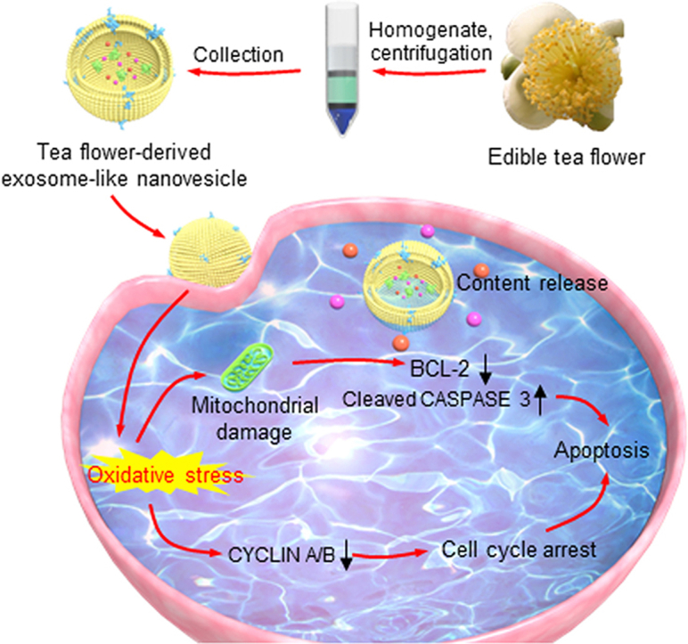
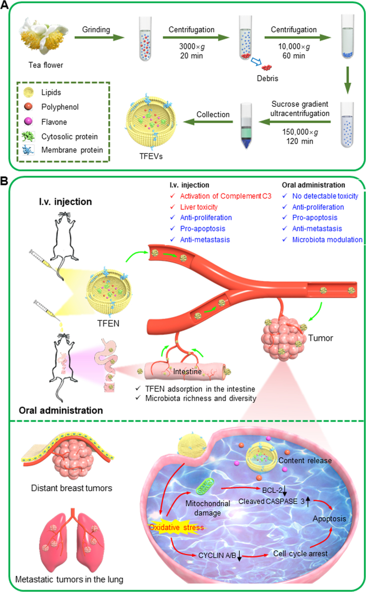
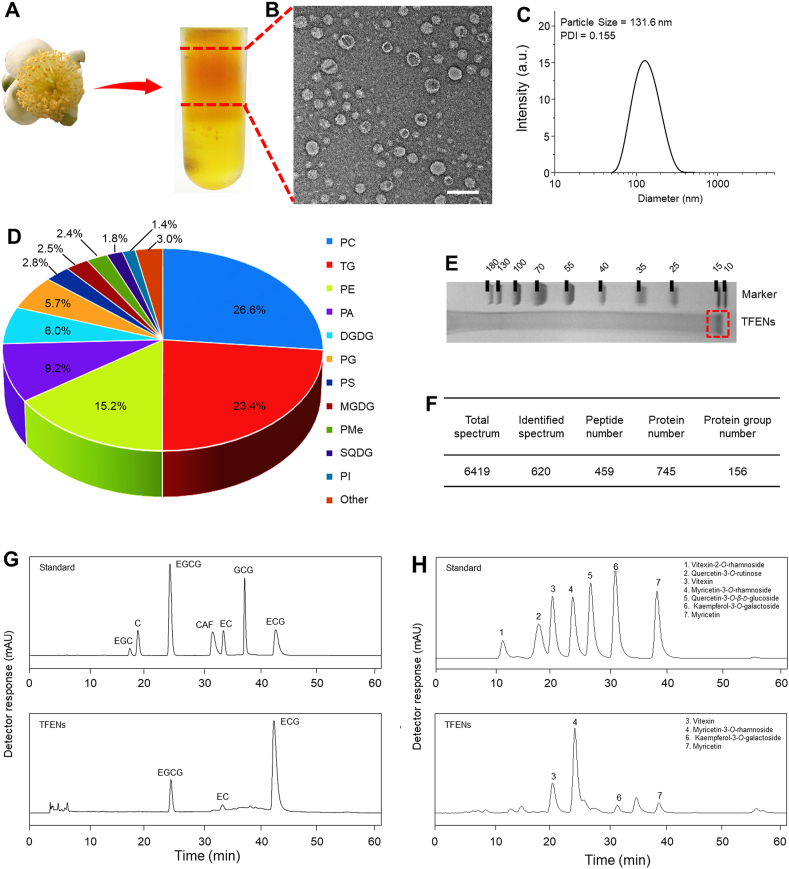
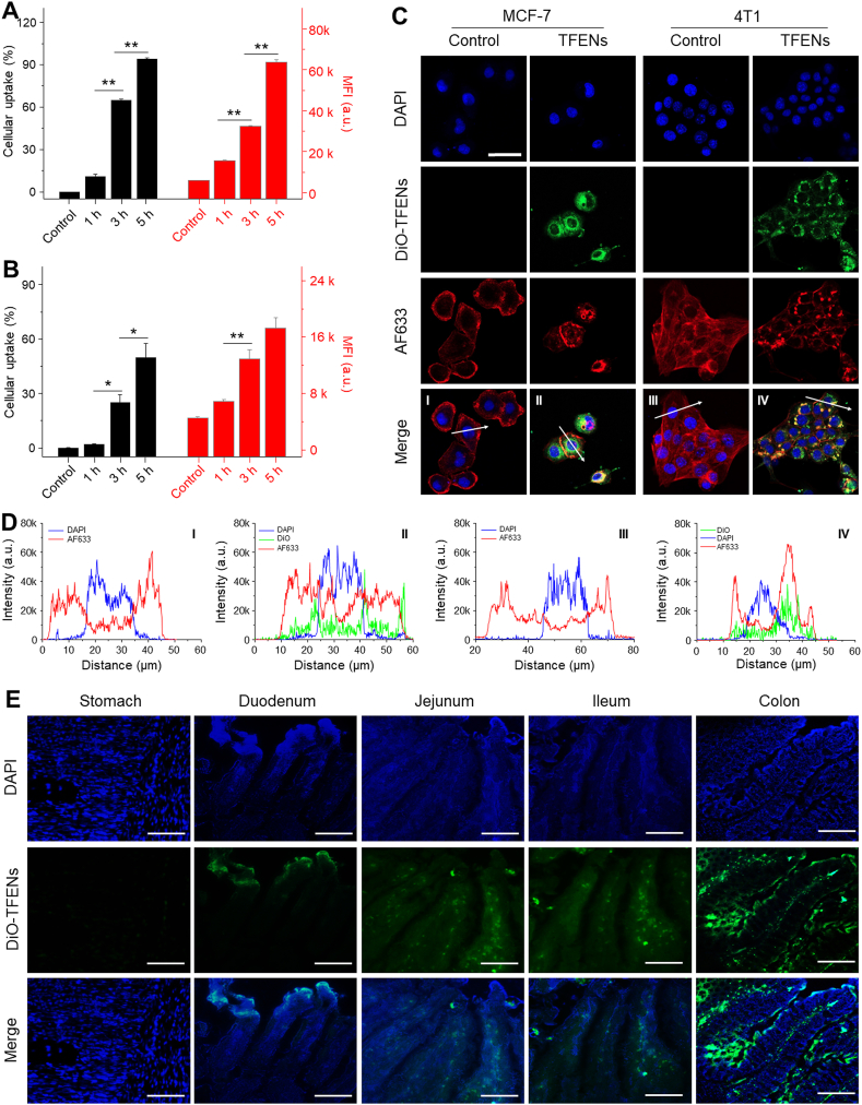
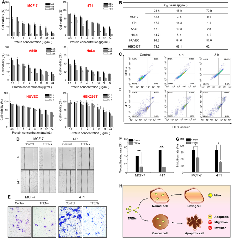
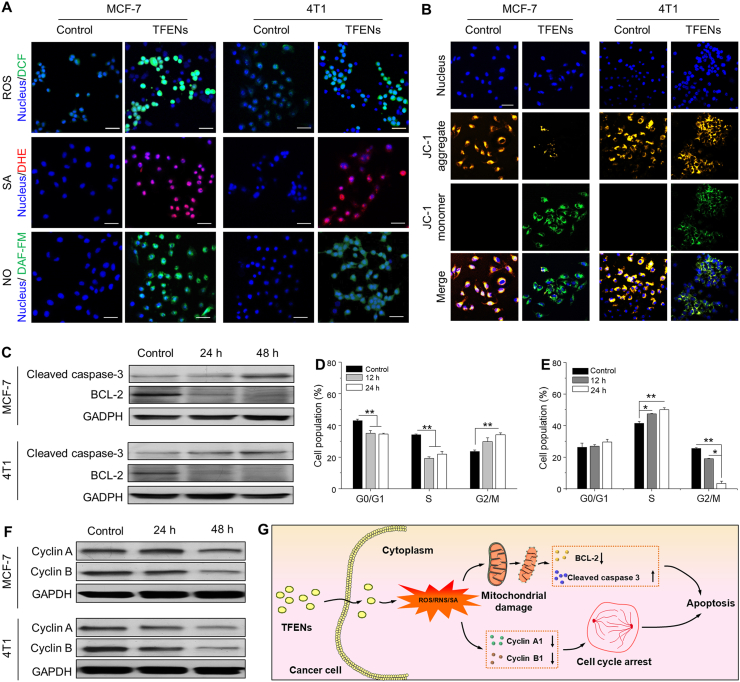
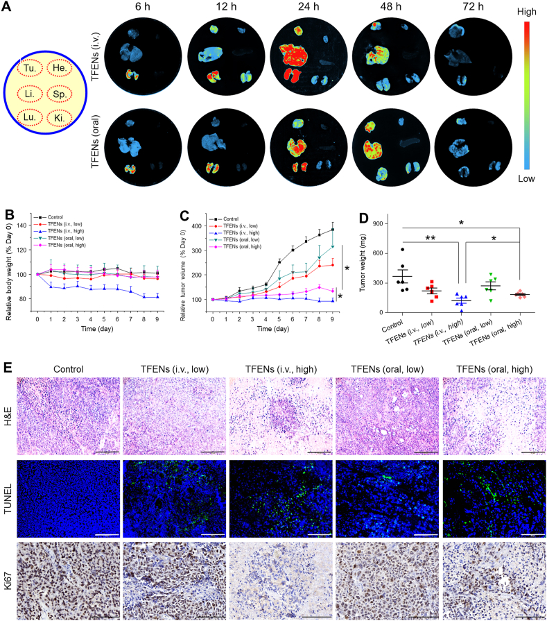
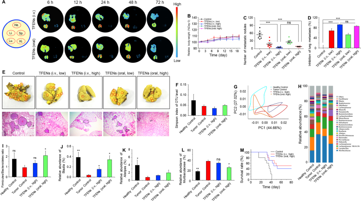
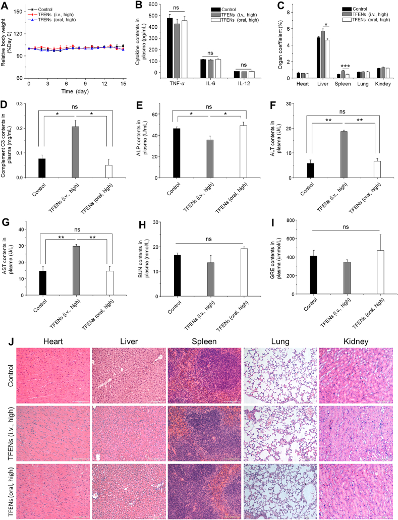
Although several artificial nanotherapeutics have been approved for practical treatment of metastatic breast cancer, their inefficient therapeutic outcomes, serious adverse effects, and high cost of mass production remain crucial challenges. Herein, we developed an alternative strategy to specifically trigger apoptosis of breast tumors and inhibit their lung metastasis by using natural nanovehicles from tea flowers (TFENs). These nanovehicles had desirable particle sizes (131 nm), exosome-like morphology, and negative zeta potentials. Furthermore, TFENs were found to contain large amounts of polyphenols, flavonoids, functional proteins, and lipids. Cell experiments revealed that TFENs showed strong cytotoxicities against cancer cells due to the stimulation of reactive oxygen species (ROS) amplification. The increased intracellular ROS amounts could not only trigger mitochondrial damage, but also arrest cell cycle, resulting in the in vitro anti-proliferation, anti-migration, and anti-invasion activities against breast cancer cells. Further mice investigations demonstrated that TFENs after intravenous (i.v.) injection or oral administration could accumulate in breast tumors and lung metastatic sites, inhibit the growth and metastasis of breast cancer, and modulate gut microbiota. This study brings new insights to the green production of natural exosome-like nanoplatform for the inhibition of breast cancer and its lung metastasis via i.v. and oral routes.

Keywords: AF633, Alexa Fluor 633-labeled phalloidin; ALP, alkaline phosphatase; ALT, alanine aminotransferase; AST, aspartate aminotransferase; BUN, urea nitrogen; Breast cancer; CDK, CYCLIN-dependent kinase; CRE, creatinine; DAF-FM DA, 4-amino-5-methylamino-2′,7′-difluorofluorescein diacetate; DAPI, 4′,6-diamidino-2-phenylindole; DCFH-DA, dichloro-dihydro-fluorescein diacetate; DGDG, digalactosyl diacylglycerols; DHE, dihydroethidium; DLS, dynamic light scattering; DiO, 3,3′-dioctadecyloxacarbocyanine perchlorate; DiR, 1,1′-dioctadecyl-3,3,3′′,3′-tetramethylindotricarbocyanine iodide; EC, epicatechin; ECG, epicatechin gallate; EGCG, epigallocatechin gallate; Exosome-like nanoparticle; FBS, fetal bovine serum; GIT, gastrointestinal tract; H&E, Hematoxylin & Eosin; HPLC, high-performance liquid chromatography; Intravenous injection; LC‒MS, liquid chromatography‒mass spectrometry; MFI, mean fluorescence intensity; MGDG, monogalactosyl diacylglycerols; MTT, 3-(4,5-dimethylthiazol-2-yl)-2,5-diphenyltetrazolium bromide; Metastasis; Microbiota modulation; NO, nitrogen monoxide; NPs, nanoparticles; OUT, operational taxonomic unit; Oral administration; PA, phosphatidic acids; PBS, phosphate-buffered saline; PC, phosphatidylcholines; PDI, polydispersity index; PE, phosphatidylethanolamines; PG, phosphatidylglycerol; PI, phosphatidylinositol; PLT, platelets; PMe, phosphatidylmethanol; PS, phosphatidylserine; RBC, red blood cell; RNS, reactive nitrogen species; ROS generation; ROS, reactive oxygen species; SA, superoxide anion; SQDG, sulphoquinovosyl diylyceride; TEM, transmission electron microscopy; TFENs, exosome-like NPs from tea flowers; TG, triglyceride; TUNEL, TdT-mediated dUTP Nick-end labeling; Tea flower; WBC, white blood cell.


Figures

Image 1
Graphical abstract
Scheme 1
Scheme 1
Schematic illustration of the production process of TFENs and their sequential treatment against metastatic breast cancers. (A) Schematic diagram of the extraction and purification of TFENs. (B) Scheme of the transportation and therapeutic functions of TFENs, including the transportation of TFENs after i.v. injection and oral administration, increased oxidative stress, mitochondrial damage, cell cycle arrest, and cell apoptosis.
Figure 1
Figure 1
Identification and physicochemical characterization of TFENs. (A) TFENs were isolated and purified by differential centrifugation and sucrose gradient ultracentrifugation. (B) TEM image (scale bar = 200 nm) and (C) hydrodynamic particle size of TFENs. The main components in TFENs (D) total lipids (E) proteins in SDS-PAGE gel (F) protein summary (G) polyphenols, and (H) flavonoids. PC, phosphatidylcholines; TG, triglyceride; PE, phosphatidylethanolamines; PA, phosphatidic acids; DGDG, digalactosyl diacylglycerols; PG, phosphatidylglycerol; PS, phosphatidylserine; MGDG, monogalactosyl diacylglycerols; PMe, phosphatidylmethanol; SQDG, sulphoquinovosyl diylyceride; PI, phosphatidylinositol; EGCG, epigallocatechin gallate; EC, epicatechin; ECG, epicatechin gallate.
Figure 2
Figure 2
Uptake profiles of TFENs by cancer cells and intestinal tissues. FCM analysis of the cellular uptake profiles of DiO-labeled TFENs in (A) MCF-7 cells and (B) 4T1 cells for 1, 3, and 5 h, respectively. Each point represents the mean ± SEM (n = 3). ∗P < 0.05, ∗∗P < 0.01. ns, no significance. (C) Confocal microscopy images and (D) fluorescence distribution profiles of MCF-7 and 4T1 cells receiving the treatment of DiO-labeled TFENs (scale bar = 20 μm). (E) Distribution of TFENs in different sections of GIT (stomach, duodenum, jejunum, ileum, and colon) from the mice with the treatment of DiO-labeled TFENs at the time point of 6 h (scale bar = 50 μm).
Figure 3
Figure 3
In vitro anti-proliferation, pro-apoptotic, and anti-mobility properties of TFENs. (A) Viabilities of various cell lines with the treatment of TFENs for 24, 48, and 72 h, respectively, and (B) their corresponding IC50 values. Each point represents the mean ± SEM (n = 5). (C) Pro-apoptotic capacities of TFENs against MCF-7 cells and 4T1 cells after co-incubation for 4 and 8 h, respectively. (D, F) Migration and (E, G) invasion of MCF-7 cells and 4T1 cells with or without the treatment of TFENs for 24 h, and their corresponding quantitative results. Scale bar = 100 μm. Each point represents the mean ± SEM (n = 3). ∗P < 0.05, ∗∗P < 0.01. ns, no significance. (H) The schematic illustration of the impacts of TFENs on healthy cells and cancer cells.
Figure 4
Figure 4
Confocal microscopy images of (A) ROS, SA, NO, and (B) mitochondrial membrane potential changes in MCF-7 cells and 4T1 cells after the treatment of TFENs for 4 h (scare bar: 50 μm). (C) Western blot analysis of cleaved caspase-3 and BCL-2 in MCF-7 cells and 4T1 cells receiving the treatment of TFENs for 24 and 48 h, respectively. Cell population profiles of (D) MCF-7 and (E) 4T1 cells in various cell cycle phases after co-incubation for 12 and 24 h, respectively. Each point represents the mean ± SEM (n = 3). ∗P < 0.05, ∗∗P < 0.01. ns, no significance). (F) Western blot analysis of cyclin A and cyclin B in MCF-7 cells and 4T1 cells receiving the treatment of TFENs for 24 and 48 h, respectively. (G) Schematic illustration of the pro-apoptotic mechanism of TFENs against cancer cells.
Figure 5
Figure 5
In vivo bio-distribution and anti-cancer activities of TFENs based on a subcutaneous xenograft breast tumor model. (A) In vivo bio-distribution of DiR-loaded TFENs in tumor, heart, liver, spleen, lung, and kidney at different time points (6, 12, 24, 48, and 72 h). (B) Relative body weight variations and (C) tumor volume variations over 9 days after the treatment of TFENs via i.v. injection and oral route. (D) Tumor weights on day 9. (E) Histological analysis of resected tumor sections from different treatment groups through H&E staining, TUNEL assay, and Ki-67 staining (scale bar = 100 μm). Each point represents the mean ± SEM (n = 6). ∗P < 0.05, ∗∗P < 0.01. ns, no significance.
Figure 6
Figure 6
In vivo bio-distribution and anti-metastasis activities of TFENs based on lung metastasis model of breast cancer. (A) In vivo bio-distribution of DiR-loaded TFENs in heart, liver, spleen, lung, and kidney at different time points (6, 12, 24, 48, and 72 h). (B) Relative body weight variations over 9 days after the treatment of TFENs via i.v. injection and oral route. (C) Total numbers of metastatic tumor nodules and (D) lung metastasis inhibition profiles of TFENs at the end of experiments. (E) Representative images and H&E staining of the lungs from different treatment groups on day 19 (scale bar = 100 mm). Microbiome analysis of the fecal samples. (f) α-Diversities were presented by box plot of the Simpson indexes. (G) Principle component analysis (PCA) plots of gut microbiota. (H) Gut microbiota compositions in different treatment groups at the genus level. (I–L) Relative abundances of the typical beneficial bacteria and harmful bacteria in different treatment groups. (M) Survival rates for each group receiving different treatments. Each point represents the mean ± SEM (n = 8). ∗P < 0.05, ∗∗P < 0.01. ns, no significance.
Figure 7
Figure 7
In vivo biosafety evaluation of different treatment approaches. (A) Body weight variations (B) pro-inflammatory cytokine levels (C) organ coefficients (D) complement C3 (E) ALP (F) ALT (G) AST (H) BUN, and (I) GRE in plasma collected from the mice with the treatment of TFENs via i.v. and oral routes. (J) Histological analysis of the five main organs from the mice with the treatment of TFENs via i.v. and oral routes (scale bar = 100 μm). Each point represents the mean ± SEM (n = 3). ∗P < 0.05, ∗∗P < 0.01. ns, no significance.

References

    1. Tang Y., Wang Y., Kiani M.F., Wang B. Classification, treatment strategy, and associated drug resistance in breast cancer. Clin Breast Cancer. 2016;16:335–343. - PubMed
    1. Kodack D.P., Askoxylakis V., Ferraro G.B., Fukumura D., Jain R.K. Emerging strategies for treating brain metastases from breast cancer. Cancer Cell. 2015;27:163–175. - PMC - PubMed
    1. Xiao Y., Cong M., Li J., He D., Wu Q., Tian P., et al. Cathepsin C promotes breast cancer lung metastasis by modulating neutrophil infiltration and neutrophil extracellular trap formation. Cancer Cell. 2021;39:423–437 e7. - PubMed
    1. Poonia N., Lather V., Pandita D. Mesoporous silica nanoparticles: a smart nanosystem for management of breast cancer. Drug Discov Today. 2018;23:315–332. - PubMed
    1. Panagi M., Voutouri C., Mpekris F., Papageorgis P., Martin M.R., Martin J.D., et al. TGF-beta inhibition combined with cytotoxic nanomedicine normalizes triple negative breast cancer microenvironment towards anti-tumor immunity. Theranostics. 2020;10:1910–1922. - PMC - PubMed