. 2022 Jan 12;7(2):146-161.
doi: 10.1016/j.jacbts.2021.11.005. eCollection 2022 Feb.

H2 Inhibits the Formation of Neutrophil Extracellular Traps

Affiliations

H2 Inhibits the Formation of Neutrophil Extracellular Traps

Kohsuke Shirakawa et al. JACC Basic Transl Sci. .

Abstract

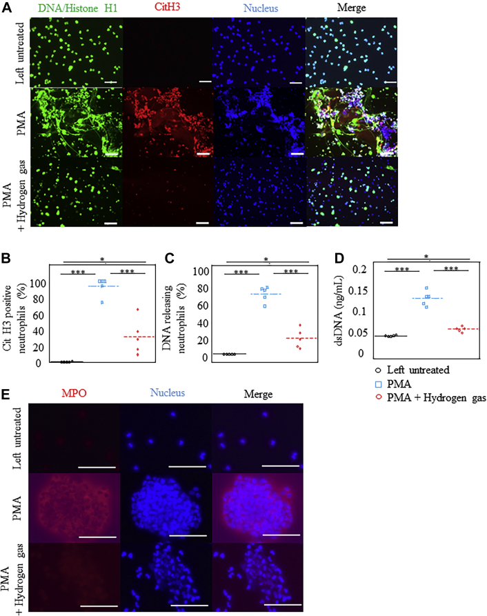
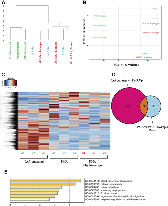
Neutrophil extracellular traps (NETs) contribute to inflammatory pathogenesis in numerous conditions, including infectious and cardiovascular diseases, and have attracted attention as potential therapeutic targets. H2 acts as an antioxidant and has been clinically and experimentally proven to ameliorate inflammation. This study was performed to investigate whether H2 could inhibit NET formation and excessive neutrophil activation. Neutrophils isolated from the blood of healthy volunteers were stimulated with phorbol-12-myristate-13-acetate (PMA) or the calcium ionophore A23187 in H2-exposed or control media. Compared with control neutrophils, PMA- or A23187-stimulated human neutrophils exposed to H2 exhibited reduced neutrophil aggregation, citrullination of histones, membrane disruption by chromatin complexes, and release of NET components. CXCR4high neutrophils are highly prone to NETs, and H2 suppressed Ser-139 phosphorylation in H2AX, a marker of DNA damage, thereby suppressing the induction of CXCR4 expression. H2 suppressed both myeloperoxidase chlorination activity and production of reactive oxygen species to the same degree as N-acetylcysteine and ascorbic acid, while showing a more potent ability to inhibit NET formation than these antioxidants do in PMA-stimulated neutrophils. Although A23187 formed NETs in a reactive oxygen species-independent manner, H2 inhibited A23187-induced NET formation, probably via direct inhibition of peptidyl arginine deiminase 4-mediated histone citrullination. Inhalation of H2 inhibited the formation and release of NET components in the blood and bronchoalveolar lavage fluid in animal models of lipopolysaccharide-induced sepsis (mice and aged mini pigs). Thus, H2 therapy can be a novel therapeutic strategy for NETs associated with excessive neutrophil activation.

Keywords: BAL, bronchoalveolar lavage; CVD, cardiovascular disease; CitH3, citrullinated histone H3; H2; HOCl, hypochlorous acid; LPS, lipopolysaccharide; MI, myocardial infarction; MPO, myeloperoxidase; NAC, N-acetyl-L-cysteine; NET, neutrophil extracellular trap; PA, pulmonary artery; PADI4, peptidyl arginine deiminase 4; PMA, phorbol-12-myristate-13-acetate; ROS, reactive oxygen species; dsDNA, double-stranded DNA; neutrophil extracellular traps; phorbol-12-myristate-13-acetate.


Conflict of interest statement

This research was funded by Japanese Society for the Promotion of Science (JSPS) Grant-in-Aid for Scientific Research (B) grant 18H02812 (2018-2020) and grants from Doctors Man Co, Ltd (to MS); and JSPS Grant-in Aid for Young Scientists grant JP18K15197 (2018–2020), Grant in-Aid for JSPS Fellows grant JP19J00583H (2018-2020). The funders had no role in the design and conduct of the study; collection, management, analysis, and interpretation of the data; preparation, review, or approval of the manuscript; or decision to submit the manuscript for publication. Drs Kobayashi and Sano have received advisory fees and research fees from Doctors Man Co, Ltd. Dr Sano has received advisory fees and research fees from Taiyo Nippon Sanso; is the registered inventor of the following patents jointly filed by Keio University and Taiyo Nippon Sanso: hydrogen mixed gas supply device for medical purposes (patent number: 5631524), medicinal composition for improving prognosis after restart of patient’s own heartbeat, and medicinal composition for improving and/or stabilizing circulatory dynamics after onset of hemorrhagic shock; and 3 other patents (whose names are translated into English): pharmaceutical compositions for reducing weight loss after organ harvesting (joint application with Keio University and Taiyo Nippon Sanso), method for generating organ preservation solution containing hydrogen and organ preservation solution containing hydrogen (joint application with Keio University and Doctors Man, application number PCT/JP2019/045790). All other authors have reported that they have no relationships relevant to the contents of this paper to disclose.

Figures

None
Graphical abstract
Figure 1
Figure 1
H2 Inhibited the Formation of NETs by Human Neutrophils Neutrophils purified from the blood of healthy volunteers were left untreated or stimulated with phorbol-12-myristate-13-acetate (PMA) in either a H2-exposed medium or control medium for 1 hour. (A) Microscopic images depicting the fluorescence of DNA/histone H1 (Alexa Fluor 488, green), citrullination of histone 3 (CitH3) (citrulline R2 + R8 + R17; Alexa Fluor 594, red), and 4′,6-diamidino-2-phenylindole (blue) in cultured neutrophils. Original magnification ×20. Bars = 100 μm. (B,C) Percentage of CitH3-positive or DNA-releasing neutrophils (n = 5). (D) Quantification of double-stranded DNA (dsDNA) released by activated neutrophils into the culture medium (n = 6). (E) Microscopic images depicting the fluorescence of myeloperoxidase (MPO) (Alexa Fluor 594, red) and 4′,6-diamidino-2-phenylindole (blue) in cultured neutrophils. Original magnification ×120. Bars = 50 μm). ∗P < 0.05; ∗∗∗P < 0.001. NET = neutrophil extracellular trap.
Figure 2
Figure 2
Bioinformatic Analysis of RNA-Seq Data From Human Neutrophils Neutrophils purified from the blood of healthy volunteers were stimulated with phorbol-12-myristate-13-acetate (PMA) in either a H2-exposed medium or control medium for 1 hour. (A) The dendrogram depicting hierarchical clustering of all RNA-sequencing (RNA-seq) samples. (B) Principal component (PC) analysis plot of all RNA-seq samples. Replicates of the same cell type are indicated by the same color, as shown in the legend. (C) Heat map of gene expression profiles shown with gene-wise (row) and sample-wise (column) clustering dendrograms. The color corresponds to the square root of standardized transcripts per million values by row, as shown in the legends on the upper left. (D) Venn diagram showing the overlap between genes up-regulated on PMA treatment (untreated vs PMA) with those down-regulated on subsequent H2 administration (PMA vs PMA + H2). (E) Gene Ontology (GO) enrichment analysis of biological processes for the 26 overlapping genes.
Figure 3
Figure 3
H2 Ameliorated the DNA Damage in Human Neutrophils Neutrophils purified from the blood of healthy volunteers were stimulated with PMA in either a H2-exposed medium or control medium for 1 hour. (A) Representative flow-cytometric analysis of phospho-histone H2A.X (Ser-139) indicating neutrophils. (B) Percentage of SYTOX+ neutrophils (n = 3). (C) Flow cytometric analysis of CXCR4+ cells in neutrophils (n = 3). (D,E) Flow cytometric analysis of SYTOX+ CXCR4+ cells in neutrophils (n = 3). (F) Microscopic images depicting the fluorescence of MPO (Alexa Fluor 594, red), DNA/histone H1 (Alexa Fluor 488, green), and 4′,6-diamidino-2-phenylindole (blue) in cultured neutrophils. Original magnification ×20. Bars = 100 μm. (G) Mean fluorescence intensity of MPO in neutrophils (n = 5) measured using the particle analysis function of the BZ-H1C software (KEYENCE). (H) Quantification of dsDNA released by activated neutrophils into the culture medium at the indicated time points (n = 6). Flow cytometric analysis was performed in n ≥ 3 independent experiments. ∗P < 0.05; ∗∗P < 0.01; ∗∗∗P < 0.001. AU = arbitrary unit; other abbreviations as in Figure 1.
Figure 4
Figure 4
H2 Reduced the Production of Hydroxy Radicals and HOCl in PMA-Stimulated Human Neutrophils Neutrophils purified from the blood of healthy volunteers were stimulated with PMA in either a H2-exposed medium or control medium for 1 hour. (A) Time course of MPO chlorination activity (n = 6). (B) Hypochlorous acid (HOCl) generation, as assessed by MPO chlorination activity (n = 6). (C) Latex beads were added to cultured human neutrophils for 1 hour and were analyzed by flow cytometry. Representative flow cytometric analysis of latex bead–positive cells in human neutrophils among different groups is depicted, indicating phagocytosis. (D) Chemotaxis assay of cultured human neutrophils. N-formylmethionyl-leucyl-phenylalanine–treated neutrophils were allowed to migrate through a porous membrane filter for 3 hours. The mean fluorescence intensity of migrated neutrophils among different groups is depicted (n = 3). ∗∗∗P < 0.001. NS = not significant; RFU = relative fluorescence unit; other abbreviations as in Figures 1 and 3.
Figure 5
Figure 5
Inhalation of H2 Inhibited NET Formation in BAL Fluid of Mice (A) Microscopic images depicting DNA/histone H1 (Alexa Fluor 488, green), MPO (Alexa Fluor 594, red), and nuclei (4′,6-diamidino-2-phenylindole, blue) in the bronchoalveolar lavage (BAL) fluid from mice that were intratracheally administered with lipopolysaccharide (E coli serotype O55:B5) and kept in cages perfused with either H2 or control gas. Original magnification ×20. Bar = 100 μm. (B) Quantification of the percentage of DNA-releasing neutrophils in the BAL fluid of each group (n = 5). (C) Quantification of CitH3 in the BAL fluid of each group (n = 5). (D) Quantification of dsDNA released in the BAL fluid of each group (n = 5). (E) Microscopic images depicting DNA/histone H1 (Alexa Fluor 488, green), MPO (Alexa Fluor 594, red), and nuclei (4′,6-diamidino-2-phenylindole, blue) in the blood of mice that were intratracheally administered with lipopolysaccharide and kept in cages perfused with either H2or control gas. Original magnification ×20. Bar = 100 μm. (F) Quantification of the percentage of DNA-releasing neutrophils in the blood of each group (n = 5). (G) Quantification of CitH3 in the plasma of each group (n = 5). (H) Quantification of dsDNA released in the plasma of each group (n = 5). ∗P < 0.05; ∗∗P < 0.01, ∗∗∗P < 0.001. Abbreviations as in Figure 1.
Figure 6
Figure 6
Inhalation of H2 Inhibited NET Formation in the PAs of Micro Mini Pigs (A) Microscopic images depicting DNA/histone H1 (Alexa Fluor 488, green) and nuclei (4′,6-diamidino-2-phenylindole, blue) in purified neutrophils isolated from the pulmonary artery (PA) of micro mini pigs administered with 20 μg/kg lipopolysaccharide intravenously with the inhalation of H2 or control gas. Original magnification ×20. Bars = 100 μm. (B) Quantification of the percentage of DNA-releasing neutrophils (n = 3). (C) Quantification of dsDNA released in the blood from the PA of each group (n = 3). (D) Representative immunofluorescence images of lung sections from micro mini pigs administered with 20 μg/kg lipopolysaccharide intravenously with the inhalation of H2 or control gas. Microscopic images depicting the fluorescence of CitH3 (Alexa Fluor 594, red), DNA/histone H1 (Alexa Fluor 488, green), and 4′,6-diamidino-2-phenylindole (blue). Original magnifications ×40 and ×120, respectively. Bars = 100 μm. (E) Number of CitH3-positive cells in low power field (LPF) (n = 5). ∗∗P < 0.01; ∗∗∗P < 0.001. Abbreviations as in Figure 1.

References

    1. Brinkmann V., Reichard U., Goosmann C., et al. Neutrophil extracellular traps kill bacteria. Science. 2004;303(5663):1532–1535. - PubMed
    1. Grover S.P., Mackman N. Neutrophils, NETs, and immunothrombosis. Blood. 2018;132(13):1360–1361. - PubMed
    1. Middleton E.A., He X.Y., Denorme F., et al. Neutrophil extracellular traps contribute to immunothrombosis in COVID-19 acute respiratory distress syndrome. Blood. 2020;136(10):1169–1179. - PMC - PubMed
    1. Song C., Li H., Li Y., et al. NETs promote ALI/ARDS inflammation by regulating alveolar macrophage polarization. Exp Cell Res. 2019;382(2):111486. - PubMed
    1. Stiel L., Mayeur-Rousse C., Helms J., Meziani F., Mauvieux L. First visualization of circulating neutrophil extracellular traps using cell fluorescence during human septic shock-induced disseminated intravascular coagulation. Thromb Res. 2019;183:153–158. - PubMed