. 2022 Apr 29;50(2):649-663.
doi: 10.1042/BST20210886.

A guide into the world of high-resolution 3D imaging: the case of soft X-ray tomography for the life sciences

Affiliations

A guide into the world of high-resolution 3D imaging: the case of soft X-ray tomography for the life sciences

Chidinma Adanna Okolo. Biochem Soc Trans. .

Abstract

In the world of bioimaging, every choice made determines the quality and content of the data collected. The choice of imaging techniques for a study could showcase or dampen expected outcomes. Synchrotron radiation is indispensable for biomedical research, driven by the need to see into biological materials and capture intricate biochemical and biophysical details at controlled environments. The same need drives correlative approaches that enable the capture of heterologous but complementary information when studying any one single target subject. Recently, the applicability of one such synchrotron technique in bioimaging, soft X-ray tomography (SXT), facilitates exploratory and basic research and is actively progressing towards filling medical and industrial needs for the rapid screening of biomaterials, reagents and processes of immediate medical significance. Soft X-ray tomography at cryogenic temperatures (cryoSXT) fills the imaging resolution gap between fluorescence microscopy (in the hundreds of nanometers but relatively accessible) and electron microscopy (few nanometers but requires extensive effort and can be difficult to access). CryoSXT currently is accessible, fully documented, can deliver 3D imaging to 25 nm resolution in a high throughput fashion, does not require laborious sample preparation procedures and can be correlated with other imaging techniques. Here, we present the current state of SXT and outline its place within the bioimaging world alongside a guided matrix that aids decision making with regards to the applicability of any given imaging technique to a particular project. Case studies where cryoSXT has facilitated a better understanding of biological processes are highlighted and future directions are discussed.


Conflict of interest statement

The author declares that there are no competing interests associated with this manuscript.

Figures

Figure 1.
Figure 1.. An exemplar full-field transmission X-ray microscope end station at beamline B24, Diamond Light Source.
(a) A view of the outer components of the microscope's end station. (b) Different components of the end station including optics and sample storage. (c) Minimum intensity projection of a 3D reconstructed tomogram derived from tilt series captured by the X-ray detector- Image courtesy of Dr Angus Wann (University of Oxford, U.K.). Arrows are pointing at different cellular components: green- endoplasmic reticulum, yellow- mitochondria, blue- cristae, red- nuclear envelope, purple- lipid droplets, orange- nucleus. Scale bar = 1 µm.
Figure 2.
Figure 2.. Imaging workflow at the correlative cryo-imaging beamline B24 at the U.K. national synchrotron, Diamond Light Source.
This platform takes samples from the preparatory stage through mapping and imaging using fully correlated SXT and super-resolution fluorescence 3D cryo-imaging leading to content-rich data collection, automated processing (provided that there are appropriate fiducial markers) and information harvest.
Figure 3.
Figure 3.. Data collection strategy for SXT.
(a) A 7 × 7 2D mosaic collected with reference applied and background subtracted. (b) A single projection of the area boxed blue in (a) during tilt series data acquisition. (c) A slice of a reconstructed 3D tomogram of the same area in (b), showing a junction between two cells. Scale bars: (a) 10 µm (b) and (c) 1 µm. Arrows are pointing at different cellular components: green- endoplasmic reticulum, yellow- mitochondria, blue- cristae, red- nuclear envelope, purple- lipid droplets, orange- cell membrane. Cyt = cytoplasm, Nu = nucleus. Image courtesy of Dr Nina Vyas (Diamond Light Source, U.K.).
Figure 4.
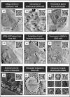
Figure 4.. Exemplar studies emanating from the imaging platform at B24.
(a) Cytotoxic T-cells producing attack particles [50]. (b) Human osteosarcoma cells (U2OS cells) showing the localization of filamentous actin with vesicles as well as the location and communications between other microstructures [49]. (c) Loss of mechanical integrity of erythrocyte membrane in the final stages of Plasmodium falciparum egress [51]. (d) SARS-CoV2 egress from Vero cells [47]. (e) Tracking of the intracellular pathway of reoviruses shows the formation of viral factories [22]. (f) Localization of reoviruses in multivesicular bodies [22]. (g) The selective cellular uptake and localization of photo-activatable anti-cancer compounds [48]. (h) Distribution trends of Chlamydiae populations in intracellular inclusions in infected human cells [53]. (i) Changes in mitochondria morphology due to an organoiridium photosensitiser [52]. Scale bars = 1 µm. N = nucleus, Cyt = cytoplasm, LD = lipid droplet, MVBs = multivesicular bodies, ER = endoplasmic reticulum, EB = elementary bodies, IM = inclusion membrane, mito = mitochondria. Images were used with permission.
Figure 5.
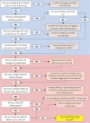
Figure 5.. Decision tree leading to cryoSXT use based on project requirements and availability of resources.
The section boxed in blue highlights sample/project-specific parameters while the section boxed in red denotes technology-related decisions.

References

    1. Abu-Siniyeh, A. and Al-Zyoud, W. (2020) Highlights on selected microscopy techniques to study zebrafish developmental biology. Lab. Anim. Res. 36, 12 10.1186/s42826-020-00044-2 - DOI - PMC - PubMed
    1. Fleming, N. (2019) The microscopic advances that are opening big opportunities in cell biology. Nature 575, S91–S94 10.1038/d41586-019-03650-w - DOI - PubMed
    1. Eggeling, C. (2018) Advances in bioimaging—challenges and potentials. J. Phys. D: Appl. Phys. 51, 040201 10.1088/1361-6463/aaa259 - DOI
    1. Fenster, A. (2002) A TRENDS guide to imaging technologies. Trends Biotechnol. 20, S1–S2 10.1016/S0167-7799(02)02024-3 - DOI - PubMed
    1. Rupprecht, J.-F., Martinez-Marrades, A., Zhang, Z., Changede, R., Kanchanawong, P. and Tessier, G. (2017) Trade-offs between structural integrity and acquisition time in stochastic super-resolution microscopy techniques. Opt. Express 25, 23146–23163 10.1364/OE.25.023146 - DOI - PubMed

MeSH terms