. 2022 Jul;110(7):1401-1415.
doi: 10.1002/jbm.a.37381. Epub 2022 Mar 8.

Photocurable antimicrobial silk-based hydrogels for corneal repair

Affiliations

Photocurable antimicrobial silk-based hydrogels for corneal repair

Inês A Barroso et al. J Biomed Mater Res A. 2022 Jul.

Abstract

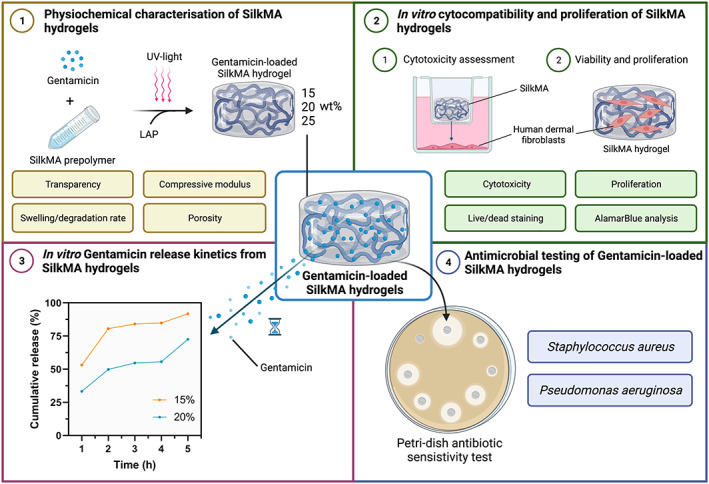
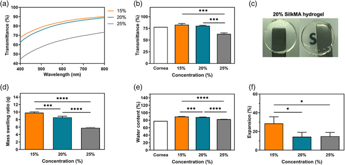
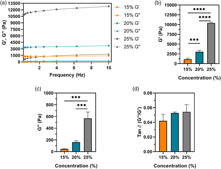
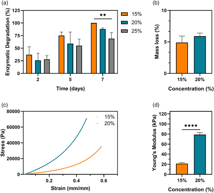
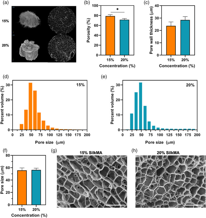
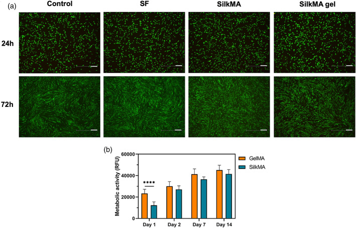
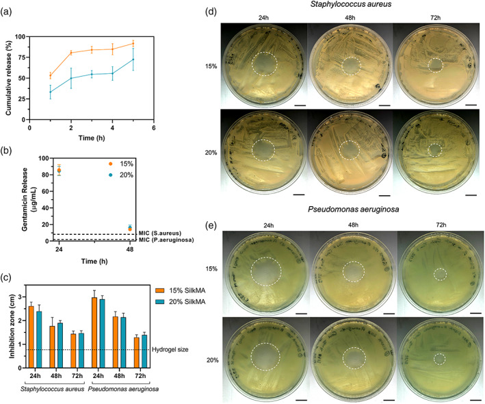
Corneal transplantation is the current gold standard treatment to restore visual acuity to patients with severe corneal diseases and injuries. Due to severe donor tissue shortage, efforts to develop a corneal equivalent have been made but the challenge remains unmet. Another issue of concern in ocular surgery is the difficult instillation and fast drainage of antibiotic ocular eye drops as bacterial infections can jeopardize implant success by delaying or impairing tissue healing. In this study, we developed antimicrobial silk-based hydrogels that have the potential to be photoactivated in situ, fully adapting to the corneal injury shape. Gentamicin-loaded methacrylated-silk (SilkMA) hydrogels were prepared within minutes using low UV intensity (3 mW/cm2 ). SilkMA gels provided a Young's modulus between 21 and 79 kPa together with a light transmittance spectrum and water content (83%-90%) similar to the human cornea. Polymer concentration (15%-25%) was found to offer a tool for tailoring the physical properties of the hydrogels. We confirmed that the methacrylation did not affect the material's in vitro degradation and biocompatibility by observing fibroblast adhesion and proliferation. Importantly, agar diffusion tests showed that the synthesized hydrogels were able to inhibit Staphylococcus aureus and Pseudomonas aeruginosa growth for 72 h. These characteristics along with their injectability and viscoelasticity demonstrate the potential of SilkMA hydrogels to be applied in several soft tissue engineering fields. As such, for the first time we demonstrate the potential of photocurable antimicrobial SilkMA hydrogels as a novel biomaterial to facilitate corneal regeneration.

Keywords: antimicrobial; cornea; regenerative medicine; silk fibroin.


Figures

FIGURE 1
FIGURE 1
Experimental outline investigating the properties of gentamicin‐loaded SilkMA hydrogels. (1) Photocrosslinking of 15%, 20%, and 25% gentamicin‐loaded SilkMA hydrogels. The transparency in the visible range, swelling, in vitro degradation, porosity/pore size, compressive Young's modulus, and rheological properties of the SilkMA hydrogels were studied. (2) In vitro cytocompatibility of gentamicin‐loaded SilkMA hydrogels. Firstly, the cytocompatibility of SF and SilkMA prepolymer solutions and SilkMA hydrogels was assessed. Then, the ability of human dermal fibroblast (HDF) cells to attach and proliferate on SilkMA hydrogels was studied and compared with GelMA hydrogels. (3) In vitro gentamicin drug release from SilkMA hydrogels. The cumulative drug release of gentamicin from SilkMA hydrogels and gentamicin concentration in the media were calculated. (4) Antimicrobial testing of gentamicin‐SilkMA hydrogels. The antimicrobial activity of SilkMA hydrogels against two of the most common causes of ocular infections, Staphylococcus aureus and Pseudomonas aeruginosa was assessed in vitro
FIGURE 2
FIGURE 2
SilkMA hydrogels transparency (n = 3): (A) transmittance profiles, (B) average optical transmittance in the visible range, and (C) optical clarity. Hydrogel diameter = 10 mm. Swelling characteristics of SilkMA hydrogels in PBS at 32°C (n = 3): (D) swelling ratio (q), (E) water content (%), and (F) expansion (%). Data presented as mean value ± SD (*p<.0001)
FIGURE 3
FIGURE 3
Rheological properties of SilkMA hydrogels (n = 3): (A) representative frequency sweep, (B) storage modulus (G′), (C) loss modulus (G″) and (D) damping factor (tan ∂). Data presented as mean value ± SD (*p<.05, **p<.01, ***p<.001, ****p<.0001)
FIGURE 4
FIGURE 4
(A) In vitro degradation of SilkMA hydrogels incubated in 1 U/ml collagenase II at 32°C for 2, 5, and 7 days (n = 5). (B) Measurement of the macromers not crosslinked in the hydrogel network after photocrosslinking (mass loss, %) (n = 3). Cyclic compressive mechanical testing of SilkMA hydrogels (n = 3): (C) representative compressive strain‐stress curves and (D) Young's moduli. Data presented as mean value ± SD (*p<.05, **p<.01, ***p<.001, ****p<.0001)
FIGURE 5
FIGURE 5
Morphology and microstructure analysis of SilkMA hydrogels after freeze drying (n = 3): (A) 3D micro‐CT reconstruction and 2D images (slices) of SilkMA scaffolds. Diameter = 7 mm. Quantitative analysis of porosity by micro‐CT: (B) total porosity, (C) mean pore thickness, (D,E) pore size distribution in the hydrogels, and (F) mean pore size. Representative SEM images of freeze‐dried SilkMA scaffolds: (G) 15% SilkMA, and (H) 20% SilkMA. Scale bar: 150 μm. Data presented as mean value ± SD (*p<.05, **p<.01, ***p<.001, ****p<.0001)
FIGURE 6
FIGURE 6
Cytocompatibility study: (A) Live/Dead images of HDFs seeded on tissue culture well‐plates with media (control) and SF/SilkMA solutions and SilkMA gels in inserts (n = 3). Scale bar = 200 μm. (B) Quantification of the metabolic activity of HDFs seeded on SilkMA and GelMA hydrogels by AlamarBlue assay 1, 2, 7, and 14 days after cell seeding (n = 5). Data presented as mean value ± SD (*****p<.0001)
FIGURE 7
FIGURE 7
In vitro properties of gentamicin‐loaded SilkMA hydrogels (n = 5): (A) Cumulative gentamicin drug release from SilkMA scaffolds after 1, 2, 3, 4, and 5 h. (B) Gentamicin concentration in the media after 24 and 48 h. Zone of inhibition study (n = 3): (C) Average zone of inhibition of SilkMA hydrogels after 24, 48, and 72 h. (D) Staphylococcus aureus, and (E) Pseudomonas aeruginosa. Scale bar = 1.5 cm

References

    1. Li L, Lu C, Wang L, et al. Gelatin‐based photocurable hydrogels for corneal wound repair. ACS Appl Mater Interfaces. 2018;10:13283‐13292. - PubMed
    1. Chen F, Le P, Fernandes‐Cunha GM, Heilshorn SC, Myung D. Bio‐orthogonally crosslinked hyaluronate‐collagen hydrogel for suture‐free corneal defect repair. Biomaterials. 2020;255:120176. - PMC - PubMed
    1. Kilic Bektas C, Burcu A, Gedikoglu G, Telek HH, Ornek F, Hasirci V. Methacrylated gelatin hydrogels as corneal stroma substitutes: in vivo study. J Biomater Sci Polym Ed. 2019;30:1803‐1821. - PubMed
    1. Fernandes‐Cunha GM, Chen KM, Chen F, et al. In situ‐forming collagen hydrogel crosslinked via multi‐functional PEG as a matrix therapy for corneal defects. Sci Rep. 2020;10:1‐13. - PMC - PubMed
    1. Duarte Campos DF, Rohde M, Ross M, et al. Corneal bioprinting utilizing collagen‐based bioinks and primary human keratocytes. J Biomed Mater Res ‐ Part A. 2019;107:1945‐1953. - PubMed