Review
. 2022 May:218:109007.
doi: 10.1016/j.exer.2022.109007. Epub 2022 Mar 4.

Immune regulation of the ocular surface

Affiliations
Review

Immune regulation of the ocular surface

Yihe Chen et al. Exp Eye Res. 2022 May.

Abstract

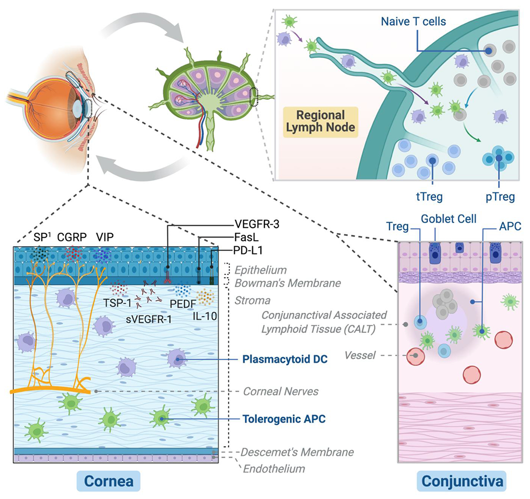
Despite constant exposure to various environmental stimuli, the ocular surface remains intact and uninflamed while maintaining the transparency of the cornea and its visual function. This 'immune privilege' of the ocular surface is not simply a result of the physical barrier function of the mucosal lining but, more importantly, is actively maintained through a variety of immunoregulatory mechanisms that prevent the disruption of immune homeostasis. In this review, we focus on essential molecular and cellular players that promote immune quiescence in steady-state conditions and suppress inflammation in disease-states. Specifically, we examine the interactions between the ocular surface and its local draining lymphoid compartment, by encompassing the corneal epithelium, corneal nerves and cornea-resident myeloid cells, conjunctival goblet cells, and regulatory T cells (Treg) in the context of ocular surface autoimmune inflammation (dry eye disease) and alloimmunity (corneal transplantation). A better understanding of the immunoregulatory mechanisms will facilitate the development of novel, targeted immunomodulatory strategies for a broad range of ocular surface inflammatory disorders.

Keywords: Alloimmunity; Autoimmunity; Immune regulation; Inflammation; Ocular surface.


Conflict of interest statement

Conflict of interest:

R.D. is consultant to Novartis, GSK, and Kala and holds equity in Claris Biotherapeutics, Aramis Biosciences, GelMEDIX, and Kera Therapeutics. Massachusetts Eye and Ear owns intellectual property related to blockade of substance P in ocular immunoinflammatory diseases.

Figures

Figure 1.
Figure 1.. The major cells and molecules promoting ocular surface immune homeostasis.
(1) Cornea: Epithelium constitutively expresses an array of immunoregulatory factors to suppress inflammatory cells and inhibit angiogenesis, including membrane-bound programmed death ligand-1 (PD-L1), Fas ligand (FasL), and vascular endothelial growth factor receptor-3 (VEGFR-3), as well as secreted pigment epithelial-derived factor (PEDF), thrombospondin-1 (TSP-1), soluble VEGFR-1 (sVEFGR-1), and IL-10. The tolerogenic antigen-presenting cells (APC) and plasmacytoid dendritic cells (DC) primarily residing in the stroma are critically involved in the induction of immune tolerance at both ocular surface and regional lymph nodes, and plasmacytoid DC additionally contribute to corneal nerve integrity and avascularity. The intense innervation of the cornea is essential for ocular surface barrier function and epithelial health with the support from the physiological level of substance P (SP) expressed by the nerves (1Excessive SP, on the contrary, promotes ocular surface inflammation and tissue damage). Other cornea nerve-derived neuropeptides, including vasoactive intestinal peptide (VIP) and calcitonin gene-related peptide (CGRP) exert important anti-inflammatory functions. (2) Conjunctiva: The specialized goblet cells in the epithelium are critical to maintaining tear film stability by secreting soluble mucins. It promotes ocular surface immune tolerance by inducing tolerogenic APC. In addition, conjunctiva-associated lymphoid tissue (CALT), which harbors regulatory T cells (Treg) is involved in the protective mucosal immune regulation. (3) Regional lymph nodes: Ocular surface adaptive immunity is primarily regulated in the local eye-draining lymph nodes by the specialized immunoregulatory cells – Treg, which include the larger thymus-derived Treg (tTreg) population and the smaller but more potent peripheral induced Treg (pTreg) population after antigen stimulation by tolerogenic APC or plasmacytoid DC.

References

    1. Bron AJ, de Paiva CS, Chauhan SK, Bonini S, Gabison EE, Jain S, et al. TFOS DEWS II pathophysiology report. Ocul Surf 2017;15:438–510. 10.1016/j.jtos.2017.05.011. - DOI - PubMed
    1. Cursiefen C Immune privilege and angiogenic privilege of the cornea. Chem Immunol Allergy 2007;92:50–7. 10.1159/000099253. - DOI - PubMed
    1. Hamrah P, Liu Y, Zhang Q, Dana MR. The corneal stroma is endowed with a significant number of resident dendritic cells. Investig Ophthalmol Vis Sci 2003;44:581–9. 10.1167/iovs.02-0838. - DOI - PubMed
    1. Hattori T, Chauhan SK, Lee H, Ueno H, Dana R, Kaplan DH, et al. Characterization of langerin-expressing dendritic cell subsets in the normal cornea. Investig Ophthalmol Vis Sci 2011;52:4598–604. 10.1167/iovs.10-6741. - DOI - PMC - PubMed
    1. Hattori T, Takahashi H, Dana R. Novel insights into the immunoregulatory function and localization of dendritic cells. Cornea 2016;35:S49–54. 10.1097/ICO.0000000000001005. - DOI - PMC - PubMed

Publication types