Review
. 2022 Sep;32(3):625-641.
doi: 10.1007/s00062-022-01144-3. Epub 2022 Mar 8.

Magnetic Resonance Imaging in Primary Progressive Multiple Sclerosis Patients : Review

Affiliations
Review

Magnetic Resonance Imaging in Primary Progressive Multiple Sclerosis Patients : Review

Malgorzata Siger. Clin Neuroradiol. 2022 Sep.

Abstract

The recently developed effective treatment of primary progressive multiple sclerosis (PPMS) requires the accurate diagnosis of patients with this type of disease. Currently, the diagnosis of PPMS is based on the 2017 McDonald criteria, although the contribution of magnetic resonance imaging (MRI) to this process is fundamental. PPMS, one of the clinical types of MS, represents 10%-15% of all MS patients. Compared to relapsing-remitting MS (RRMS), PPMS differs in terms of pathology, clinical presentation and MRI features. Regarding conventional MRI, focal lesions on T2-weighted images and acute inflammatory lesions with contrast enhancement are less common in PPMS than in RRMS. On the other hand, MRI features of chronic inflammation, such as slowly evolving/expanding lesions (SELs) and leptomeningeal enhancement (LME), and brain and spinal cord atrophy are more common MRI characteristics in PPMS than RRMS. Nonconventional MRI also shows differences in subtle white and grey matter damage between PPMS and other clinical types of disease. In this review, we present separate diagnostic criteria, conventional and nonconventional MRI specificity for PPMS, which may support and simplify the diagnosis of this type of MS in daily clinical practice.

Keywords: Diagnosis of multiple sclerosis; Differential diagnosis.


Conflict of interest statement

The author declares that there is no conflict of interest.

Figures

Fig. 1
Fig. 1
Active MS lesion (Gd+ lesion) in a primary progressive MS patient. a Axial T1-weighted image after contrast administration in a PPMS patient shows a gadolinium-enhancing lesion (white arrow) in juxtacortical white matter. b Corresponding section of axial fluid attenuated inversion recovery (FLAIR) image with hyperintense white matter lesion (arrowhead) in the same location
Fig. 2
Fig. 2
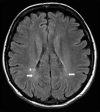
Focal white matter lesions in three PPMS patients. a Axial fluid attenuated inversion recovery (FLAIR) images with only a few small, focal white matter lesions located in deep white matter in right and left brain hemisphere (white arrows), b axial FLAIR image with punctate lesion in right deep white matter (white arrow), c axial FLAIR image with small, focal lesion located in deep white matter of right brain hemisphere (white arrow) and in periventricular white matter (white arrowhead) of left brain hemisphere
Fig. 3
Fig. 3
Brain MRI scans in PPMS patients who are similar to those seen in relapse–remitting multiple sclerosis (RRMS) and secondary progressive multiple sclerosis (SPMS). Brain MRI scans of PPMS patients with different MRI presentations a FLAIR image, axial plane. Multiple focal lesions were located in the periventricular, deep and juxtacortical white matter. This MRI is similar to a typical MRI scan observed with RRMS. b Fluid attenuated inversion recovery (FLAIR) image, axial plane. Focal and confluent white matter lesions were located in the periventricular white matter; some lesions located in the juxtacortical regions were similar to those seen in SPMS. Signs of brain atrophy are also visible
Fig. 4
Fig. 4
Cortical/juxtacortical lesions in a patient with PPMS. MRI scan in a patient with PPMS. a Proton-density (PD)-weighted image in the axial plane. Nonvisible cortical lesion in the right parietal region. b Fluid attenuated inversion recovery (FLAIR) image, axial plane. A poorly visible cortical lesion in the right parietal region (white arrow). c Double inversion recovery (DIR) image of a clearly visible lesion in the same location (white arrowhead)
Fig. 5
Fig. 5
Black holes in four PPMS patients. ad Axial T1-weighted spin-echo images with hypointense lesions (black holes) in deep and periventricular white matter (white arrows), eh. Axial fluid attenuated inversion recovery (FLAIR) images with corresponding hyperintense lesions in the same locations
Fig. 6
Fig. 6
Dirty appearing white matter (DAWM) in a patient with PPMS. Fluid attenuated inversion recovery (FLAIR) image in the axial plane. Around the posterior horns of the lateral ventricles, ill-defined areas of increased signal intensity are visible (white arrow). Right periventricular hyperintense focal lesions are also visible (asterisk)
Fig. 7
Fig. 7
Jacobian analysis and SEL candidates [63] (Reprinted by permission of SAGE Publication). ab An axial slice of linearly coregistered reference and follow-up T1-weighted scans. c The reference scan with a regular grid overlaid. di The nonlinearly deformed image in c is shown to match the follow-up scan, and dii an enlarged lesion area of the deformation field. e The Jacobian determinant is shown as a heat map, where blue represents local contraction and red represents local expansion. The Jacobian determinant represents the local percent volume change at each voxel after application of the nonlinear deformation that warps (a) to match (b). f An axial slice of a reference T2-weighted scan with overlaid T2 lesion segmentation. g The Jacobian determinant within reference T2 lesions. h Initial SEL candidate boundaries based on JE1. i Refined SEL candidate boundaries based on JE2. JE Jacobian expansion, SEL slowly expanding/evolving lesion
Fig. 8
Fig. 8
MRI of focal lesions in the spinal cord of a patient with PPMS. a T2-weighted, turbo spin-echo image, sagittal plane. Hyperintense focal lesions at the C1 C3, C4, C5 spinal cord levels (white arrows). b Corresponding T2-weighted turbo spin-echo image, axial section at C1 spinal cord level. Hyperintense focal lesions at right side of spinal cord. c T1-weighted, turbo spin-echo, sagittal plane without signal abnormalities and corresponding axial section at C1 spinal cord level (d) also without signal abnormalities. e T1-weighted images after contrast administration, sagittal plane. Gd+ lesion at C1 spinal cord level (white arrow) and axial section at the same spinal cord level (f) with Gd+ lesion (arrowhead)
Fig. 9
Fig. 9
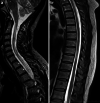
Diffuse abnormalities in the spinal cord of a patient with PPMS. An MRI scan of the spinal cord in a patient with PPMS. a Cervical spinal cord, T2-weighted image, sagittal plane. Diffuse, hyperintense signal abnormalities in the whole cervical spinal cord. b Thoracic spinal cord; T2-weighted image in the sagittal plane. Characteristic features of diffuse abnormalities with heterogeneous signals at multiple levels and spinal cord atrophy are visible
Fig. 10
Fig. 10
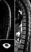
MRI of spinal cord atrophy in a patient with PPMS. Spinal cord atrophy in a patient with PPMS a T2-weighted image in the sagittal plane; marked atrophy of the thoracic spinal cord segments with visible hyperintense signal abnormalities. b T2-weighted image in the axial plane of thoracic segment (Th9–Th10), marked atrophy with hyperintense signal reflects demyelinating lesion. c T1-weighted image, sagittal plane in the same patient with marked atrophy in thoracic spinal cord

References

    1. Montalban X, Hauser SL, Kappos L, Arnold DL, Bar-Or A, Comi G, de Seze J, Giovannoni G, Hartung HP, Hemmer B, Lublin F, Rammohan KW, Selmaj K, Traboulsee A, Sauter A, Masterman D, Fontoura P, Belachew S, Garren H, Mairon N, Chin P, Wolinsky JS, ORATORIO Clinical Investigators Ocrelizumab versus placebo in primary progressive multiple sclerosis. N Engl J Med. 2017;19:209–220. doi: 10.1056/NEJMoa1606468. - DOI - PubMed
    1. Wolinsky JS, Arnold DL, Brochet B, Hartung HP, Montalban X, Naismith RT, Manfrini M, Overell J, Koendgen H, Sauter A, Bennett I, Hubeaux S, Kappos L, Hauser SL. Long-term follow-up from the ORATORIO trial of ocrelizumab for primary progressive multiple sclerosis: a post-hoc analysis from the ongoing open-label extension of the randomised, placebo-controlled, phase 3 trial. Lancet Neurol. 2020;19:998–1009. doi: 10.1016/S1474-4422(20)30342-2. - DOI - PubMed
    1. Hawker K, O’Connor P, Freedman MS, Calabresi PA, Antel J, Simon J, Hauser S, Waubant E, Vollmer T, Panitch H, Zhang J, Chin P, Smith CH, OLYMPUS trial group Rituximab in patients with primary progressive multiple sclerosis: results of arandomized double-blind placebo-controlled multicenter trial. Ann Neurol. 2009;66:460–471. doi: 10.1002/ana.21867. - DOI - PubMed
    1. Thompson AJ, Banwell BL, Barkhof F, Carroll WM, Coetzee T, Comi G, Correale J, Fazekas F, Filippi M, Freedman MS, Fujihara K, Galetta SL, Hartung HP, Kappos L, Lublin FD, Marrie RA, Miller AE, Miller DH, Montalban X, Mowry EM, Sorensen PS, Tintoré M, Traboulsee AL, Trojano M, Uitdehaag BMJ, Vukusic S, Waubant E, Weinshenker BG, Reingold SC, Cohen JA. Diagnosis of multiple sclerosis: 2017revisions of the McDonald criteria. Lancet Neurol. 2018;17:162–173. doi: 10.1016/S1474-4422(17)30470-2. - DOI - PubMed
    1. Filippi M, Rocca MA, Ciccarelli O, De Stefano N, Evangelou N, Kappos L, Rovira A, Sastre-Garriga J, Tintorè M, Frederiksen JL, Gasperini C, Palace J, Reich DS, Banwell B, Montalban X, Barkhof F, MAGNIMS Study Group MRI criteria for the diagnosis of multiple sclerosis: MAGNIMS consensus guidelines. Lancet Neurol. 2016;15:292–303. doi: 10.1016/S1474-4422(15)00393-2. - DOI - PMC - PubMed