. 2022 Mar 23;144(11):5087-5098.
doi: 10.1021/jacs.1c13706. Epub 2022 Mar 8.

Mechanism of Radical S-Adenosyl- l-methionine Adenosylation: Radical Intermediates and the Catalytic Competence of the 5'-Deoxyadenosyl Radical

Affiliations

Mechanism of Radical S-Adenosyl- l-methionine Adenosylation: Radical Intermediates and the Catalytic Competence of the 5'-Deoxyadenosyl Radical

Maike N Lundahl et al. J Am Chem Soc. .

Abstract

Radical S-adenosyl-l-methionine (SAM) enzymes employ a [4Fe-4S] cluster and SAM to initiate diverse radical reactions via either H-atom abstraction or substrate adenosylation. Here we use freeze-quench techniques together with electron paramagnetic resonance (EPR) spectroscopy to provide snapshots of the reaction pathway in an adenosylation reaction catalyzed by the radical SAM enzyme pyruvate formate-lyase activating enzyme on a peptide substrate containing a dehydroalanine residue in place of the target glycine. The reaction proceeds via the initial formation of the organometallic intermediate Ω, as evidenced by the characteristic EPR signal with g = 2.035 and g = 2.004 observed when the reaction is freeze-quenched at 500 ms. Thermal annealing of frozen Ω converts it into a second paramagnetic species centered at giso = 2.004; this second species was generated directly using freeze-quench at intermediate times (∼8 s) and unequivocally identified via isotopic labeling and EPR spectroscopy as the tertiary peptide radical resulting from adenosylation of the peptide substrate. An additional paramagnetic species observed in samples quenched at intermediate times was revealed through thermal annealing while frozen and spectral subtraction as the SAM-derived 5'-deoxyadenosyl radical (5'-dAdo•). The time course of the 5'-dAdo• and tertiary peptide radical EPR signals reveals that the former generates the latter. These results thus support a mechanism in which Ω liberates 5'-dAdo• by Fe-C5' bond homolysis, and the 5'-dAdo• attacks the dehydroalanine residue of the peptide substrate to form the adenosylated peptide radical species. The results thus provide a picture of a catalytically competent 5'-dAdo• intermediate trapped just prior to reaction with the substrate.


Conflict of interest statement

The authors declare no competing financial interest.

Figures

Figure 1.
Figure 1.
5′-Deoxyadenosyl radical (5′-dAdo•) generated from reductive cleavage by RS enzyme and subsequent reactivity. Pathway (a) hydrogen atom abstraction from the substrate resulting in substrate radical. Pathway (b) adenosylation of a substrate via 5′-dAdo• addition to sp2 carbon.
Figure 2.
Figure 2.
Structure of the 8-mer Dha-pep.
Figure 3.
Figure 3.
RFQ and annealing of the PFL-AE/SAM/Dha-pep reaction. (A) RFQ of the reaction of PFL-AE with SAM and Dha-pep at 500 ms, 1, and 2 s shows the increasing intensity of an EPR signal characteristic of the organometallic intermediate Ω. (B) Cryogenic annealing of the 2 s RFQ sample at the temperatures and times shown results in conversion of Ω to a new radical species. The signal from this species is saturation broadened at the observation temperature of 40 K, chosen because Ω is not observable at the higher temperatures optimal for the radical. EPR conditions: microwave frequency, 9.375 GHz; modulation amplitude, 5 G, T = 40 K.
Figure 4.
Figure 4.
Normalized 70 K X-band EPR spectra (black) and simulations (red) of the Ado-Dha-pep• generated from the reaction of reduced PFL-AE, SAM, and Dha-pep at RT then freeze-quenched at ~8 s. (A) Reaction using natural abundance (N.A.) SAM and Dha-pep. (B) Reaction was carried out using D2-Dha-pep. (C) Reaction using [adenosyl-13C10,15N5]-SAM. (D) Reaction using natural abundance SAM and Dha-pep was carried out in 95% D2O. (E) Reaction using natural abundance SAM and D2-Dha-pep was carried out in 95% D2O. Total spin quantitations in (A–E) range from 11 to 20 μM. EPR conditions: microwave frequency, 9.38 GHz; modulation, 3 G, T = 70 K. Simulations were generated with Easyspin with g-tensor = [2.0047 2.0039 2.0025] and hyperfine tensors aiso(Ha) = 45 MHz, aiso(N–H) = 30 MHz, and aiso(13C(5′)) = 48 MHz.
Figure 5.
Figure 5.
Structure considered for the Ado-Dha-pep•. Left, depiction of the fixed geometry of the Ado-Dha-pep•. Center, illustration of the dihedral angle between the 2pπ orbital on Cα and the amide N–H bond for the fixed geometry observed in the PFL-AE crystal structure, as viewed down the Cα–Namide bond. Right, illustration of the dihedral angles between the 2pπ orbital on Cα and the Cβ–H and Cβ–C5′ bonds, viewed down the Cα–Cβ bond. The plane of the sp2 Cα, Namide, and Cβ is represented by a dashed line.
Figure 6.
Figure 6.
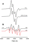
EPR spectra at 40 K of a 10 s freeze-quench sample before and after annealing reveal the presence of 5′-dAdo•. (A) The CW X-band EPR spectra were recorded at 40 K of the 10 s freeze-quench reaction of PFL-AE, SAM, and Dha-pep before and after annealing at 125 K for 15 min. (B) The difference spectrum generated by subtracting the post-anneal spectrum from the pre-anneal spectrum is shown in black, with the simulation in red. For spin quantitation, see Table S2. EPR conditions: microwave frequency, 9.38 GHz; modulation amplitude, 3 G, T = 40 K.
Figure 7.
Figure 7.
EPR spectra of freeze-quench time course samples from 8 to 18 s. (A) The CW X-band EPR spectra recorded at 40 K of reactions of PFL-AE, SAM, and Dha-pep freeze-quenched at times from 8 to 18 s. (B) Spectra in panel A were simulated to estimate contributions from the 5′-dAdo• and the Ado-Dha-pep• at each time point. For spin quantitation, see Table S4. EPR conditions: microwave frequency, 9.38 GHz; modulation amplitude, 3 G, T = 40 K.
Figure 8.
Figure 8.
Models of the Dha peptide and its adenosylated product in the PFL-AE active site from PDB 3CB8. The 4Fe–4S cluster (orange and yellow), sodium ion (purple), SAM (cyan carbons), and peptide (green carbons) are depicted in sticks with oxygen atoms colored red and nitrogen atoms colored blue. Hydrogen bonding interactions of PFL-AE to the adenosine of SAM are yellow dashed lines. (A) Representation of the Dha-pep in the active site of PFL-AE where the black dashed line indicates the 3.7 Å distance from 5′-C to Cβ. (B) Representation of the Ado-Dha-pep• in the active site of PFL-AE showing the H-bonding from the protein active site to the adenosine is retained.
Scheme 1.
Scheme 1.
Capture of a Tertiary Carbon Radical Intermediate

References

    1. Frey PA; Hegeman AD; Ruzicka FJ The radical SAM superfamily. Crit. Rev. Biochem. Mol. Biol 2008, 43, 63–88. - PubMed
    1. Broderick JB; Duffus BR; Duschene KS; Shepard EM Radical S-Adenosylmethionine Enzymes. Chem. Rev 2014, 114, 4229–4317. - PMC - PubMed
    1. Landgraf BJ; McCarthy EL; Booker SJ Radical S-adenosylmethionine enzymes in human health and disease. Annu. Rev. Biochem 2016, 85, 485–514. - PubMed
    1. Bridwell-Rabb J; Grell TAJ; Drennan CL A rich man, poor man story of S-adenosylmethionine and cobalamin revisited. Annu. Rev. Biochem 2018, 87, 555–584. - PubMed
    1. Nicolet Y Structure-function relationships of radical of radical SAM enzymes. Nat. Catal 2020, 3, 337–350.

Publication types