. 2022 Aug;77(2):344-352.
doi: 10.1016/j.jhep.2022.02.024. Epub 2022 Mar 5.

Hepatic recruitment of eosinophils and their protective function during acute liver injury

Affiliations

Hepatic recruitment of eosinophils and their protective function during acute liver injury

Long Xu et al. J Hepatol. 2022 Aug.

Abstract

Background & aims: Beyond the classical description of eosinophil functions in parasite infections and allergic diseases, emerging evidence supports a critical role of eosinophils in resolving inflammation and promoting tissue remodeling. However, the role of eosinophils in liver injury and the underlying mechanism of their recruitment into the liver remain unclear.

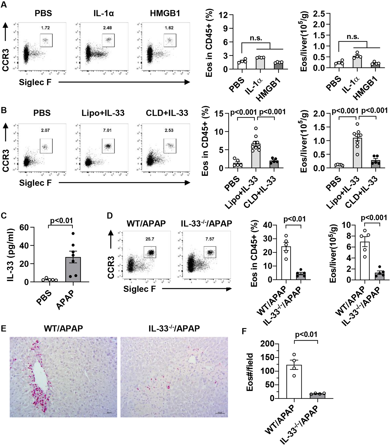
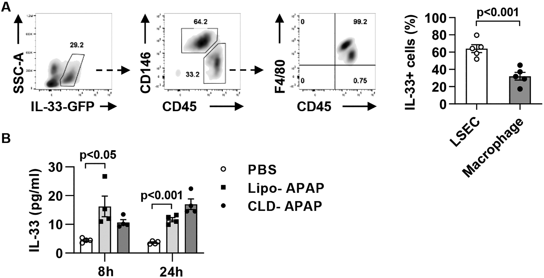
Methods: Hepatic eosinophils were detected and quantified using flow cytometry and immunohistochemical staining. Eosinophil-deficient (ΔdblGata1) mice were used to investigate the role of eosinophils in 3 models of acute liver injury. In vivo experiments using Il33-/- mice and macrophage-depleted mice, as well as in vitro cultures of eosinophils and macrophages, were performed to interrogate the mechanism of eotaxin-2 (CCL24) production.

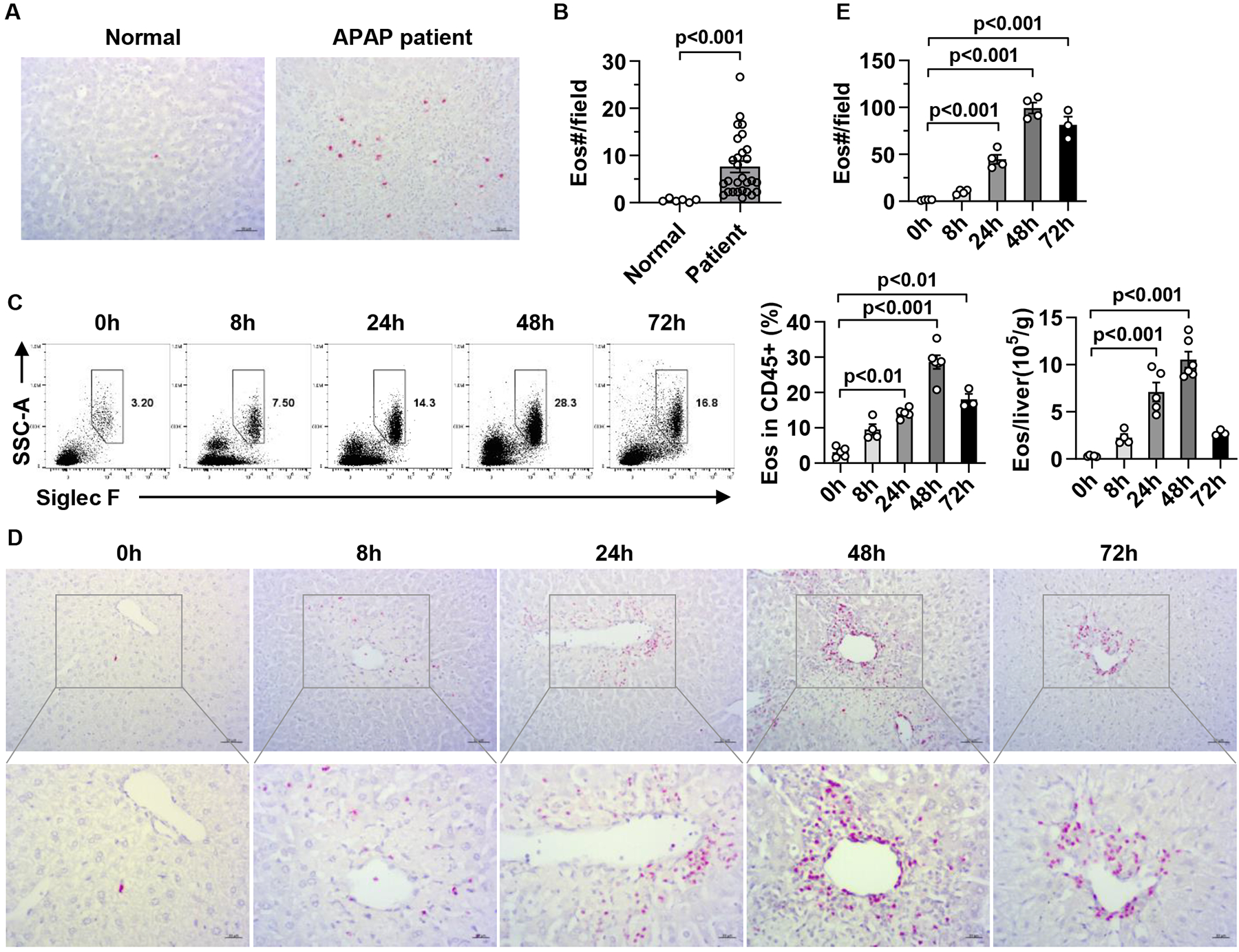
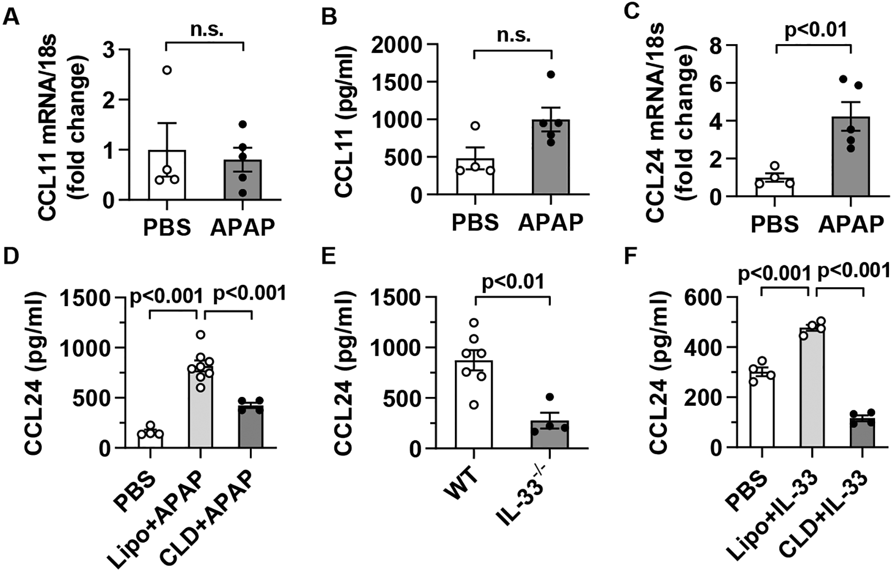
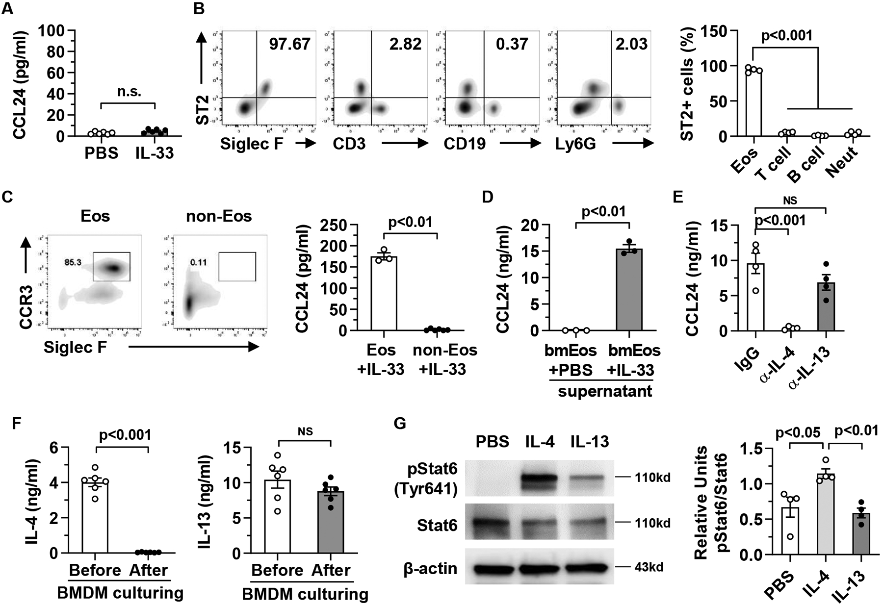
Results: Hepatic accumulation of eosinophils was observed in patients with acetaminophen (APAP)-induced liver failure, whereas few eosinophils were detectable in healthy liver tissues. In mice treated with APAP, carbon tetrachloride or concanavalin A, eosinophils were recruited into the liver and played a profound protective role. Mice deficient of macrophages or IL-33 exhibited impaired hepatic eosinophil recruitment during acute liver injury. CCL24, but not CCL11, was increased after treatment of each hepatotoxin in an IL-33 and macrophage-dependent manner. In vitro experiments demonstrated that IL-33, by stimulating IL-4 release from eosinophils, promoted the production of CCL24 by macrophages.

Conclusions: This is the first study to demonstrate that hepatic recruitment of and protection by eosinophils occur commonly in various models of acute liver injury. Our findings support further exploration of eosinophils as a therapeutic target to treat APAP-induced acute liver injury.

Lay summary: The current study unveils that eosinophils are recruited into the liver and play a protective function during acute liver injury caused by acetaminophen overdose. The data demonstrate that IL-33-activated eosinophils trigger macrophages to release high amounts of CCL24, which promotes hepatic eosinophil recruitment. Our findings suggest that eosinophils could be an effective cell-based therapy for the treatment of acetaminophen-induced acute liver injury.

Keywords: CCL24; Eosinophils; IL-33; acute liver injury; macrophages.


Conflict of interest statement

Conflict of interest The authors declare that they have no competing interests. Please refer to the accompanying ICMJE disclosure forms for further details.

Figures

Fig.1.
Fig.1.. Eosinophils accumulate in the liver after APAP treatment.
(A, B) Eosinophils in liver samples from individuals with normal livers (Normal, n=6) and patients with APAP-induced liver failure (APAP patient, n=28) were detected by IHC staining using anti-human EPX antibody (scale bar, 50μm). The numbers of eosinophils per field (776×582 pixels) were quantified. (C-E) Male C57BL/6 mice were injected i.p. with APAP. (C) The percentage and absolute number of eosinophils in the liver were measured after APAP treatment by flow cytometry (n=3–6/group). Eosinophils were identified as CD45+CD11b+SSChiSiglec F+CCR3+ cell. (D, E) Eosinophils were detected by IHC staining using anti-mouse MBP antibody and quantified (n= 3–4/group, scale bar, 50μm). Two-tailed unpaired Student’s t test was performed in B. One-way ANOVA with Tukey post hoc test was performed in C and E.
Fig. 2.
Fig. 2.. Eosinophils protect against APAP-induced liver injury.
(A to C) WT Balb/c and ΔdblGATA1 mice were i.v. injected with PBS or WT bmEos (7.5×10⁶) immediately before treatment with APAP (n=4/group). Serum ALT levels were measured, and liver necrosis (scale bars, 100μm) was evaluated and quantified at 24h after APAP treatment. (D to F) ΔdblGATA1 mice were i.v. injected with PBS or WT bmEos (7.5×10⁶) at 0h, 8h, or 12h after APAP treatment (n=3/group). Serum levels of ALT were measured and liver necrosis (scale bars, 100μm) was examined at 24h after APAP treatment. One-way ANOVA with Tukey post hoc test was performed in A, C, D, and F.
Fig. 3.
Fig. 3.. IL-33 promotes eosinophil recruitment to the liver.
(A) C57BL/6 mice were injected i.v. with PBS, IL-1α, or HMGB1 twice on one day spaced out by 8h (0.5 μg/mouse each dose). The percentage and number of hepatic eosinophils were measured by flow cytometry at 24h after first dose (n=4/group). (B) C57BL/6 mice were injected i.v. with PBS (n=5) or IL-33 (n=9) twice on one day spaced out by 8h (0.5 μg/mouse each dose). Some IL-33-treated mice were injected with CLD (n=5) to deplete macrophages or empty liposomes (Lipo) as control (n=9) at 24h prior to the initial IL-33 treatment. All mice were sacrificed at 24h after the first dose of IL-33 for the measurement of the percentage and number of hepatic eosinophils. (C) C57BL/6 mice were injected i.p. with PBS (n=5) or APAP (n=7). After 24h, serum levels of IL-33 were measured by ELISA. (D, E) WT and IL-33−/− mice were injected i.p. with APAP. After 48h, eosinophils in the liver were measured using flow cytometry (D, n=5/group) and IHC staining (E-F, n=4/group, scale bar, 50μm). Two-tailed unpaired Student’s t test was performed in A to D, and F. n.s. indicates no statistical significance.
Fig. 4.
Fig. 4.. Liver sinusoidal endothelial cells are the main source of IL-33 release during AILI.
(A) IL-33-GFP reporter mice were injected i.p. with APAP (n=5). After 24h, liver non-parenchymal cell were isolated and analyzed for GFP+ cells by flow cytometry. (B) WT C57BL/6 mice were injected i.p. with PBS (n=3) or APAP (n=8). The APAP-treated mice were divided into two groups (n=4/group) treated with empty liposomes (Lipo) or CLD to deplete macrophages 24h prior to APAP challenge. All mice were sacrificed at 24h after APAP treatment and serum levels of IL-33 were measured by ELISA. Two-tailed unpaired Student’s t test was performed in A and B.
Fig. 5.
Fig. 5.. CCL24, but not CCL11, is up-regulated in a macrophage-dependent manner during AILI.
(A-D) C57BL/6 mice were injected i.p. with PBS or APAP (n=4–5/group). After 24h, CCL11 and CCL24 mRNA expression in liver tissues (A, C) and protein levels in sera (B, D) were measured by qPCR and ELISA, respectively. Some mice were treated with CLD to deplete macrophages at 24h prior to APAP challenge. (E) WT (n=7) and IL-33−/− (n=4) mice were treated with APAP, and after 24h serum levels of CCL24 were measured. (F) C57BL/6 mice were treated with PBS or IL-33 as described in Fig. 3B. Some IL-33-treated mice were injected with CLD to deplete macrophages following the same protocol described in Fig. 3B. All mice were sacrificed at 24h after the first dose of IL-33 and serum levels of CCL24 were measured (n=4/group). Two-tailed unpaired Student’s t test was performed in A to F. n.s. indicates no statistical significance.
Fig. 6.
Fig. 6.. IL-33-stimulated eosinophils, through release of IL-4, induce CCL24 production by macrophages
(A) BMDMs (4×10⁶ cells/ml per well), generated from C57BL/6 mice, were stimulated with mouse recombinant IL-33 (20 ng/ml). After 24h, CCL24 protein levels in the supernatants were measured by ELISA. (B) Liver mononuclear cells (MNCs) were isolated from WT mice (n=4) at 24h after APAP treatment and analyzed for ST2 expression by flow cytometry. (C) From liver MNCs, isolated at 24h after APAP treatment, eosinophils (Eos) were purified by Siglec F-positive selection using MACS and the purity is around 85%. The Siglec F-negative portion of cell mixture (non-Eos) was also collected. Eos and non-Eos (1×10⁶ cells/ml per well) were cultured together with BMDMs (4×10⁶ cells/well) in presence of IL-33. After 24h co-culturing, CCL24 protein levels in the supernatants were measured by ELISA. (D) Bone marrow-derived eosinophils (bmEos, 4×10⁶ cells/ml per well) were stimulated with PBS or IL-33 (20 ng/ml). After 24h, the conditioned media (CM) were collected and used to culture BMDMs (4×10⁶ cells/ml per well) for another 24h. CCL24 protein levels in the BMDM culture supernatants were measured by ELISA. (E) The culturing system was set up as described in D with the addition of anti-IL-4 antibody or anti-IL-13 antibody in the CM collected from IL-33-stimulated bmEos. (F) The protein levels of IL-4 and IL-13 in the CM from IL-33-stimulated bmEos were measured before and after culturing of BMDMs. (G). BMDMs was treated with PBS, mouse recombinant IL-4 or IL-13 for 1h. Expression levels of phosphorylated-Stat6, Stat6, and β-actin were measured and quantified (n=4/group). Two-tailed unpaired Student’s t test was performed in A to D, and F. One-way ANOVA with Tukey post hoc test was performed in E and G. n.s. indicates no statistical significance.

References

    1. Stravitz RT, Lee WM. Acute liver failure. Lancet. 2019;394(10201):869–81. - PMC - PubMed
    1. Klion AD, Nutman TB. The role of eosinophils in host defense against helminth parasites. The Journal of allergy and clinical immunology. 2004;113(1):30–7. - PubMed
    1. Gleich GJ. Mechanisms of eosinophil-associated inflammation. The Journal of allergy and clinical immunology. 2000;105(4):651–63. - PubMed
    1. Krishack PA, Hollinger MK, Kuzel TG, Decker TS, Louviere TJ, Hrusch CL, et al. IL-33-mediated Eosinophilia Protects against Acute Lung Injury. American journal of respiratory cell and molecular biology. 2021;64(5):569–78. - PMC - PubMed
    1. Takeda K, Shiraishi Y, Ashino S, Han J, Jia Y, Wang M, et al. Eosinophils contribute to the resolution of lung-allergic responses following repeated allergen challenge. The Journal of allergy and clinical immunology. 2015;135(2):451–60. - PMC - PubMed

Publication types