

MiR-5195-3p functions as a tumor suppressor in prostate cancer via targeting CCNL1
- PMID: 35260070
- PMCID: PMC8905902
- DOI: 10.1186/s11658-022-00326-8
MiR-5195-3p functions as a tumor suppressor in prostate cancer via targeting CCNL1
Abstract
Background: Accumulating evidence indicates that miR-5195-3p exerts tumor-suppressive roles in several tumors. However, the clinical significance and biological function of miR-5195-3p in prostate cancer (PCa) have not been reported yet.
Methods: The expression levels of miR-5195-3p and Cyclin L1 (CCNL1) were determined using quantitative real-time PCR in clinical specimens and cell lines. The clinical significance of miR-5195-3p in patients with PCa was evaluated using Kaplan-Meier survival analysis and Cox regression models. Cell proliferation and cell cycle distribution were measured by CCK-8 assay and flow cytometry, respectively. The association between miR-5195-3p and CCNL1 was analyzed by luciferase reporter assay.
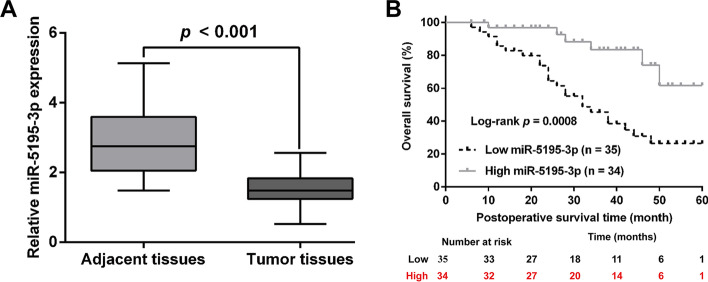
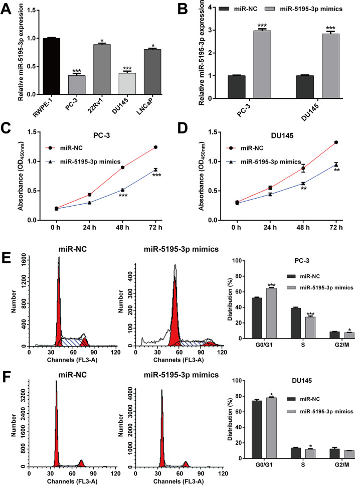
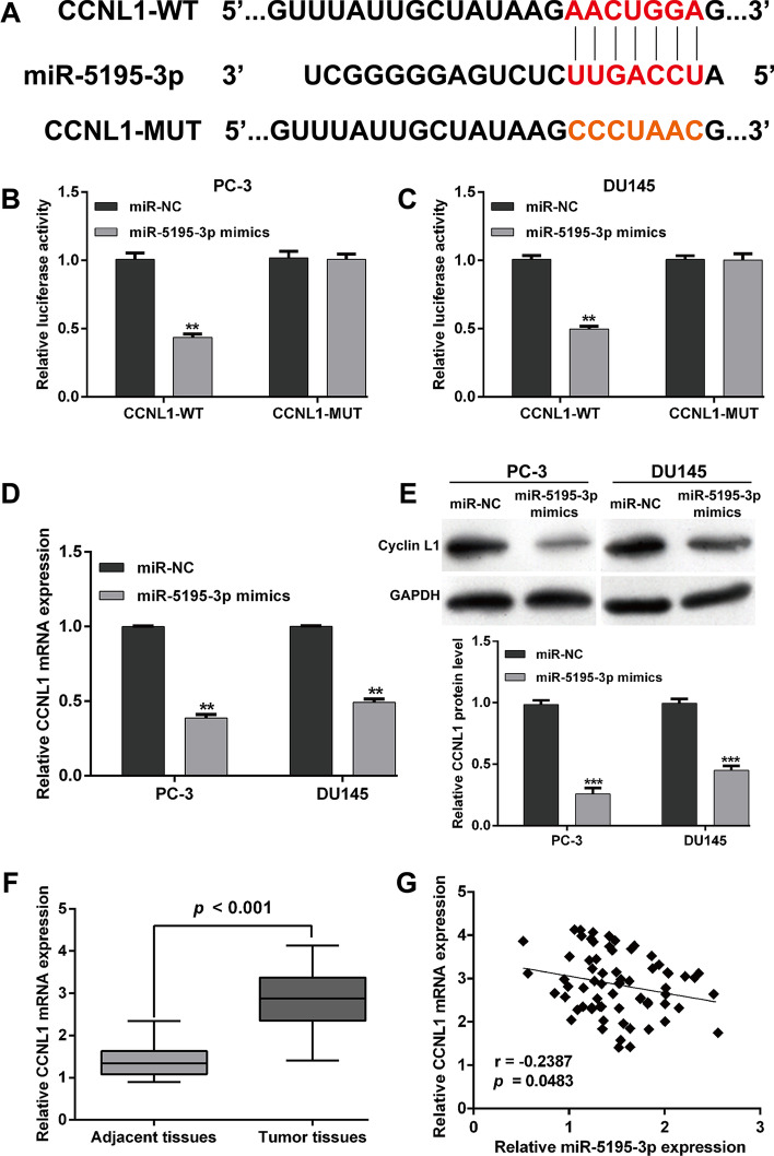
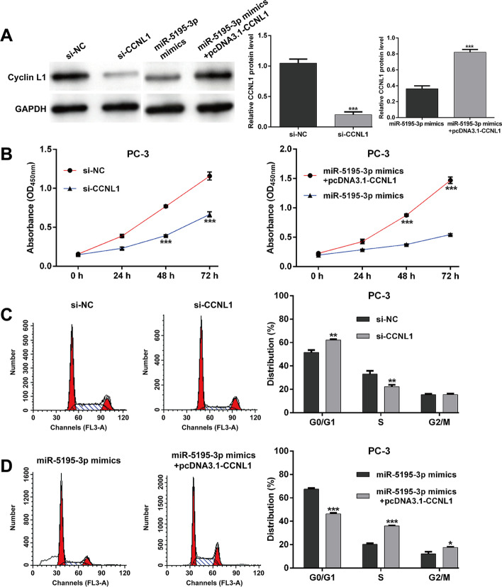
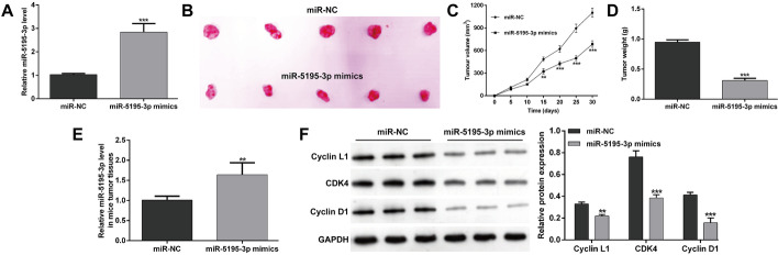
Results: MiR-5195-3p expression levels were significantly downregulated in 69 paired PCa tissues compared with matched adjacent normal tissues. The decreased miR-5195-3p expression was associated with Gleason score and TNM stage, as well as worse survival prognosis. The in vitro experiments showed that miR-5195-3p overexpression suppressed the proliferation and cell cycle G1/S transition in PC-3 and DU145 cells. Elevated miR-5195-3p abundance obviously impaired tumor formation in vivo using PC-3 xenografts. Mechanistically, CCNL1 was a direct target of miR-5195-3p in PCa cells, which was inversely correlated with miR-5195-3p in PCa tissues. Importantly, CCNL1 knockdown imitated, while overexpression reversed, the effects of miR-5195-3p overexpression on PCa cell proliferation and cell cycle G1/S transition.
Conclusions: Our data suggest that miR-5195-3p functions as a tumor suppressor by targeting CCNL1 in PCa.
Keywords: CCNL1; Proliferation; Prostate cancer; miR-5195-3p.
© 2022. The Author(s).
Conflict of interest statement
The authors declare that they have no competing interests.
Figures
Similar articles
-
MiR-5195-3p functions as a tumor suppressor by targeting RHBDD1 in ovarian cancer.Histol Histopathol. 2023 Dec;38(12):1403-1413. doi: 10.14670/HH-18-595. Epub 2023 Feb 20. Histol Histopathol. 2023. PMID: 36825753
-
MicroRNA-487a-3p functions as a new tumor suppressor in prostate cancer by targeting CCND1.J Cell Physiol. 2020 Feb;235(2):1588-1600. doi: 10.1002/jcp.29078. Epub 2019 Jul 15. J Cell Physiol. 2020. PMID: 31309555
-
Genistein up-regulates tumor suppressor microRNA-574-3p in prostate cancer.PLoS One. 2013;8(3):e58929. doi: 10.1371/journal.pone.0058929. Epub 2013 Mar 12. PLoS One. 2013. PMID: 23554959 Free PMC article.
-
MiR-1273 g-3p Promotes Malignant Progression and has Prognostic Implications in Prostate Cancer.Mol Biotechnol. 2022 Jan;64(1):17-24. doi: 10.1007/s12033-021-00384-x. Epub 2021 Aug 24. Mol Biotechnol. 2022. PMID: 34431044
-
The putative tumour suppressor miR-1-3p modulates prostate cancer cell aggressiveness by repressing E2F5 and PFTK1.J Exp Clin Cancer Res. 2018 Sep 5;37(1):219. doi: 10.1186/s13046-018-0895-z. J Exp Clin Cancer Res. 2018. PMID: 30185212 Free PMC article.
Cited by
-
MiR-5195-3p predicts clinical prognosis and represses colorectal cancer progression by targeting TLR4/MyD88 signaling.Cell Div. 2024 Oct 10;19(1):29. doi: 10.1186/s13008-024-00133-x. Cell Div. 2024. PMID: 39390599 Free PMC article.
-
n6-methyladenosine-modified circular RNA family with sequence similarity 126, member A affects cholesterol synthesis and malignant progression of prostate cancer cells by targeting microRNA-505-3p to mediate calnexin.J Cancer. 2024 Jan 1;15(4):966-980. doi: 10.7150/jca.89135. eCollection 2024. J Cancer. 2024. PMID: 38230215 Free PMC article.
-
MiR-5195-3p targets the PCBP2/PI3K/AKT pathway to inhibit melanoma cell proliferation and migration.Heliyon. 2023 Aug 19;9(9):e19227. doi: 10.1016/j.heliyon.2023.e19227. eCollection 2023 Sep. Heliyon. 2023. PMID: 37662755 Free PMC article.
-
MicroRNA-5195-3p mediated malignant biological behaviour of insulin-resistant liver cancer cells via SOX9 and TPM4.BMC Cancer. 2023 Jun 16;23(1):557. doi: 10.1186/s12885-023-11068-x. BMC Cancer. 2023. PMID: 37328795 Free PMC article.
-
Cyclin L1 participates in Adriamycin resistance and progression of osteosarcoma via PI3K/AKT-mTOR pathway.Aging (Albany NY). 2024 Jun 26;16(14):11208-11223. doi: 10.18632/aging.205972. Epub 2024 Jun 26. Aging (Albany NY). 2024. PMID: 39024509 Free PMC article.
References
Publication types
MeSH terms
Substances
LinkOut - more resources
Full Text Sources
Medical
Molecular Biology Databases
Miscellaneous
