. 2022 Mar 8;13(1):1209.
doi: 10.1038/s41467-022-28549-5.

CMTr cap-adjacent 2'-O-ribose mRNA methyltransferases are required for reward learning and mRNA localization to synapses

Affiliations

CMTr cap-adjacent 2'-O-ribose mRNA methyltransferases are required for reward learning and mRNA localization to synapses

Irmgard U Haussmann et al. Nat Commun. .

Abstract

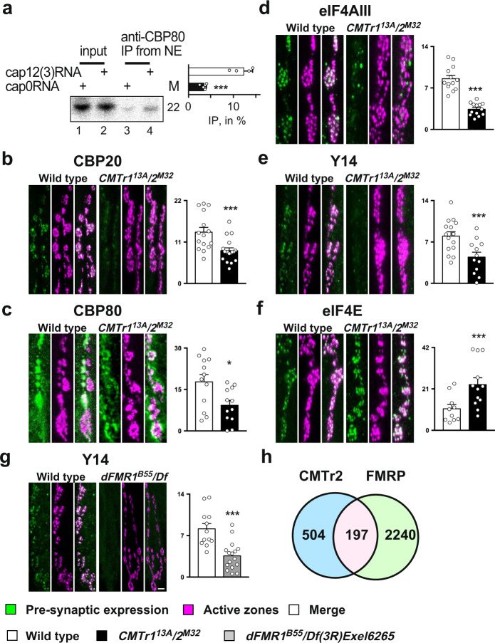
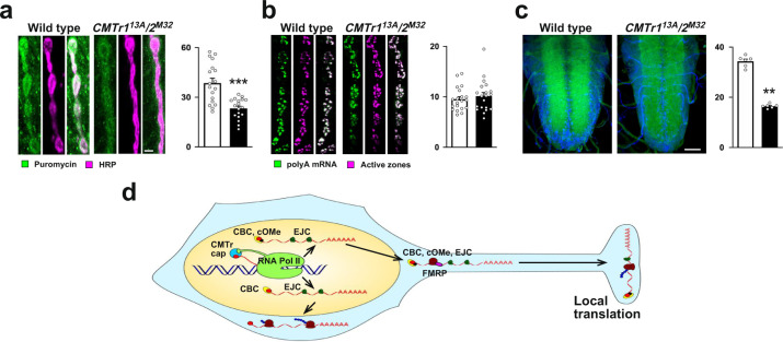
Cap-adjacent nucleotides of animal, protist and viral mRNAs can be O-methylated at the 2' position of the ribose (cOMe). The functions of cOMe in animals, however, remain largely unknown. Here we show that the two cap methyltransferases (CMTr1 and CMTr2) of Drosophila can methylate the ribose of the first nucleotide in mRNA. Double-mutant flies lack cOMe but are viable. Consistent with prominent neuronal expression, they have a reward learning defect that can be rescued by conditional expression in mushroom body neurons before training. Among CMTr targets are cell adhesion and signaling molecules. Many are relevant for learning, and are also targets of Fragile X Mental Retardation Protein (FMRP). Like FMRP, cOMe is required for localization of untranslated mRNAs to synapses and enhances binding of the cap binding complex in the nucleus. Hence, our study reveals a mechanism to co-transcriptionally prime mRNAs by cOMe for localized protein synthesis at synapses.


Conflict of interest statement

The authors declare no competing interests.

Figures

Fig. 1
Fig. 1. Analysis of CMTr1 and CMTr2 null mutants and mRNA cap 2′-O-ribose methylation in Drosophila.
a, b Genomic organization of the CMTr1 and CMTr2 loci depicting the transposons (black triangle) used to generate the deletions 13A and M32, which are null alleles. Genomic rescue fragments tagged either with hemaglutinin (HA, a) or FLAG (b) epitopes are indicated at the bottom. Primers used for validating the deletions are indicated on top of the transcript. c Validation of CMTr113A and CMTr2M32 single and double mutants by genomic PCR. The gel is representative of two biological replicates. The marker is a 100 bp DNA ladder with 500 bp indicated on top. Wild type is indicated in white, CMTr113A in light gray, CMTr2M32 in dark gray, and CMTr113A/2M32 in black. d Survival of flies to adulthood after hatching from the eggshell shown as mean ±SE (n = 3, except CMTr113A/2M32 n = 4). e Climbing activity was assessed by negative geotaxis assays shown as mean ± SE, n = 40, p = 0.005 by one-way ANOVA followed by Tukey’s test. f Bouton numbers at NMJs of muscle 13 in third instar larvae are shown as mean ± SE. n = 11, **p = 0.005 and ****p ≤ 0.0001 by one-way ANOVA followed by Tukey’s test. g Recapping of mRNA with 32PalphaGTP from adult flies of the indicated genotypes. 5′cap structures were separated on 20% denaturing polyacrylamide gels after digestion with RNAse I (lanes 5–8, right) Markers—M1: RNAse I digested 32PalphaGTP capped in vitro transcript starting with AGU and 2′-O-ribose methylated with vaccinia CMTr. M2: RNAse T1 digested 32PalphaGTP capped in vitro transcript starting with AGU. M3: RNAse I digested 32PalphaGTP capped in vitro transcript starting with AGU. Sequences of markers are shown on the left and of cap structures from adult flies are shown on the right, N: any nucleotide, *: 32P, m: methyl-group. L: single nucleotide ladder with nucleotide number indicated in white made by alkaline hydrolysis of a 5′ 32P-labeled RNA oligonucleotide. h Quantification of 5′ cap structures shown as mean ± SE. n = 3. Non-ribosemethylated cap is in black and ribose methylated in gray. i Schematic diagram of a 2D thin-layer chromatography (TLC) depicting standard and 2′-O-ribose methylated (m) phospho-nucleotides. ψ: pseudouridine. jn Representative TLCs from three replicates showing modifications of the first cap-adjacent nucleotides of S2 cells (je), adult control (k), and CMTr113A and CMTr2M32 single (l, m) and double (n) mutant females. o Quantification of the mRNA first nucleotide shown as mean ± SE from TLC (n = 5, white) and CAGEseq data (n = 8, black) from adult Drosophila and S2 cells, respectively. Source data for gels, survival to adulthood, climbing activity, bouton numbers at muscle 13, for 5′cap structures, and first nucleotide in mRNA are provided as a Source Data file.
Fig. 2
Fig. 2. mRNA cap 2′-O-ribose methylation is required for reward learning in Drosophila.
a, b Appetitive memory immediately (a) and 24 h (b) after training of control (white) and CMTr113A (light gray) and CMTrM32 (dark gray) single and double mutant (black) flies shown as mean ± SE. n = 8 for A and n = 6 for B, except CMTr113A/2M32 n = 10, p ≤ 0.006. c Rescue of the learning defect in CMTr113A; CMTr2M32 double mutant flies by genomic fragments (blue) shown as mean ± SE. n = 6, p = 0.002. d, e Rescue of the learning defect in CMTr113A; CMTr2M32 double mutant (black) flies by constitutive (d, n = 8, except UAS-CMTr2 in light gray and MB-GAL4 UASCMTr2 in blue in CMTr113A/2M32 n = 6, dark gray: MB-GAL4) or conditional (e, n = 6, except MBGSG-GAL4 UASCMTr2 in CMTr113A/2M32 n = 8) expression of CMTr2 in mushroom bodies from UAS in the absence (light gray) or presence (dark gray) of RU486 shown as mean ± SE, p ≤ 0.0001. Statistical analysis was done by one-way ANOVA followed by Dunnett’s multiple comparison test. Source data for learning and memory experiments are provided as a Source Data file.
Fig. 3
Fig. 3. Impact of mRNA cap-adjacent 2′-O-ribose methylation on gene expression and RNA stability.
a Volcano plot depicting differentially expressed genes in CMTr113A; CMTr2M32 double mutant flies compared to control flies. b Functional classification of upregulated (bottom) and downregulated (top) genes in CMTr113A; CMTr2M32 double mutant flies compared to control flies. c Incubation of monophosphorylated RNA (pRNA, gray) and capped RNA with (white) or without 2′-O-ribose methylation of cap-adjacent nucleotides (black) in nuclear (nu) and cytoplasmic (cy) extracts from S2 cells. M: length in nucleotides. The graph to the right depicts the percent undegraded RNA left after 45 min as mean ± SE (n = 5, except n = 4 for pRNA, p = 0.03 by one way ANOVA followed by Tukey’s test). Source data for gel and RNA stability values are provided as a Source Data file.
Fig. 4
Fig. 4. CMTr2 localizes to distinct sites of transcription and has a dedicated set of targets.
aj Representative images of polytene chromosomes from salivary glands from three replicates expressing CMTr1::HA (ae) or CMTr2::FLAG (fj) stained with anti-Pol II (magenta, d, i), anti-HA (green, c, h), and DNA (DAPI, blue, b, g), or merged (white, a, e, f, j). Arrowheads indicate the absence of CMTr2. Scale bars in f are 10 µm and in g are 2 µm. k Functional classification of CMTr2 CLIP targets.
Fig. 5
Fig. 5. 2′-O-ribose methylation of mRNA cap-adjacent nucleotides is required for localizing untranslated transcripts to synapses.
a Immunoprecipitation of capped RNA with or without 2′-O-ribose methylation from nuclear extracts. M: length in nucleotides. The graph to the right depicts the ratio of IP/input (n = 4, p = 0.016). bg Representative images from staining of synapses at third instar NMJs pre-synaptically expressing epitope-tagged markers with elavC155-GAL4 from UAS or stained with an antibody (CBP80) in control (left, green) and CMTr113A; CMTr2M32 double mutant larvae (bf) or dFMR1B55/Df3R)Exel6265 mutant larvae (g, right, green). The active zone of synapses was stained with nc82 (magenta). The mean ± SE of the intensity is shown on the right is arbitrary units in white for the control, in black for CMTr113A; CMTr2M32 double mutant larvae (bf) and in gray dFMR1B55/Df3R)Exel6265 mutant larvae (g) for nuclear cap-binding proteins CPB20 (b, n = 16 and 19, ***p = 0.0015) and CBP80 (c, n = 12, *p = 0.05), for nuclear exon junction complex (EJC) proteins eIF4AIII (d, n = 13, ***p = 0.005) and Y14 (e, n = 15 and12, ***p = 0.003), for the rate-limiting translation initiation factor eIF4E (f, n = 10 and 12, ***p = 0.009) and for nuclear exon junction complex (EJC) protein Y14 in control and dFMR1B55/Df3R)Exel6265 mutant larvae (g, n = 12 and 16, ***p = 0.005). The scale bar in g is 1 µm. h Overlap (pink) of CMTr2 CLIP targets (blue) with FMRP targets (green) in Drosophila. Statistical analysis was done by an unpaired t-test. Source data for gel, immunoprecipitations values, and intensity of pre-synaptic bouton stainings are provided as a Source Data file.
Fig. 6
Fig. 6. 2‘-O-ribose methylation of mRNA cap-adjacent nucleotides enhances translation at synapses.
a Staining of synapses at third instar NMJs with anti-puromycin antibodies (left, green) and with anti-HRP recognizing a neuronal epitope (right, red) after 30 min of puromycin incorporation in control and CMTr113A; CMTr2M32 double mutant larvae. The mean ± SE of the intensity is shown on the right is arbitrary units in white for the control, in black for CMTr113A; CMTr2M32 double mutant larvae (n = 18, ***p ≤ 0.001). b RNA in situ hybridization with an oligo(dT) probe to mRNA in synapses at third instar NMJs (left, green) in control and CMTr113A; CMTr2M32 double mutant larvae. The active zone of synapses was stained with nc82 (magenta, right). The mean ± SE of the intensity is shown on the right is arbitrary units in white for the control, in black for CMTr113A; CMTr2M32 double mutant larvae (n = 20). c RNA in situ hybridization with an oligo(dT) probe (green) to mRNA in the ventral nerve cord of third instar larvae (green) in control and CMTr113A; CMTr2M32 double mutant larvae counterstained with DAPI (blue). The mean ± SE of the intensity is shown on the right is arbitrary units in white for the control, in black for CMTr113A; CMTr2M32 double mutant larvae (n = 6, **p = 0.002). Scale bars in a and c are 1 µm and 50 µm, respectively. Statistical analysis was done by an unpaired t-test. d Model for the role of cap-adjacent 2′-O-ribose methylation in gene expression in neurons. EJC exon junction complex containing Y14 and eIF4AIII, CBC cap-binding complex consisting of CBP20 and 80, CMTr cap methyltransferase, FMRP Fragile X Mental Retardation Protein, cOMe cap 2′-O-ribose methylation at cap-adjacent nucleotides, ribosomes are shown as brown blobs. Source data for the intensity of pre-synaptic bouton and brain stainings are provided as a Source Data file.

Similar articles

Cited by

References

    1. Zaccara S, Ries RJ, Jaffrey SR. Reading, writing and erasing mRNA methylation. Nat. Rev. Mol. Cell Biol. 2019;20:608–624. - PubMed
    1. Frye M, Harada BT, Behm M, He C. RNA modifications modulate gene expression during development. Science. 2018;361:1346–1349. - PMC - PubMed
    1. Roignant JY, Soller M. m6A in mRNA: an ancient mechanism for fine-tuning gene expression. Trends Genet. 2017;33:380–390. - PubMed
    1. Livneh I, Moshitch-Moshkovitz S, Amariglio N, Rechavi G, Dominissini D. The m(6)A epitranscriptome: transcriptome plasticity in brain development and function. Nat. Rev. Neurosci. 2020;21:36–51. - PubMed
    1. Anreiter I, Mir Q, Simpson JT, Janga SC, Soller M. New twists in detecting mRNA modification dynamics. Trends Biotechnol. 2021;39:72–89. - PMC - PubMed

Publication types

MeSH terms