. 2022 Mar 8;13(1):1218.
doi: 10.1038/s41467-022-28828-1.

Bacterial F-type ATP synthases follow a well-choreographed assembly pathway

Affiliations

Bacterial F-type ATP synthases follow a well-choreographed assembly pathway

Khanh Vu Huu et al. Nat Commun. .

Abstract

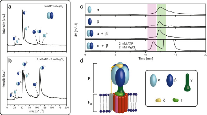
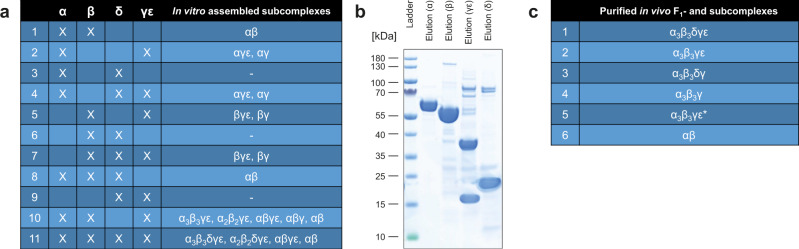
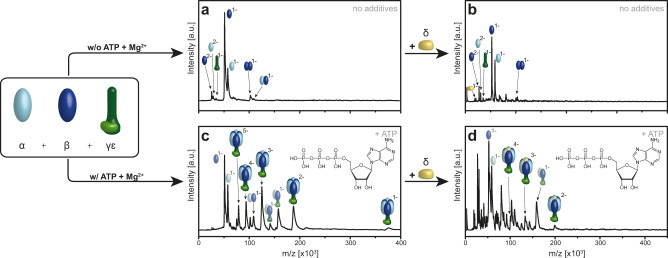
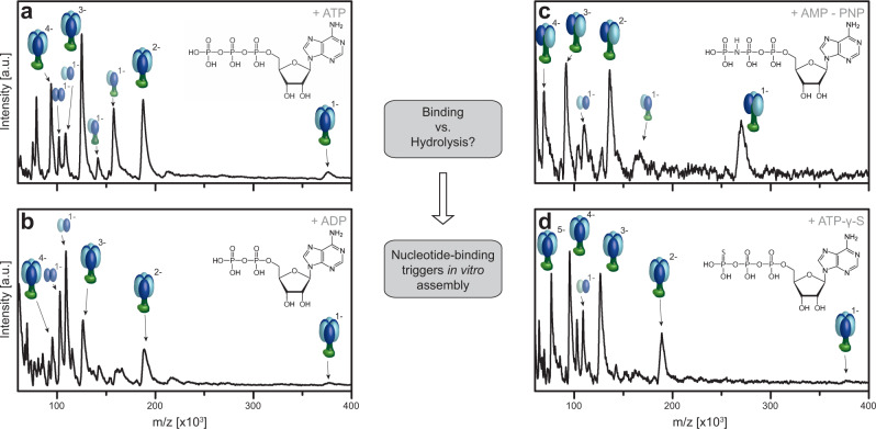
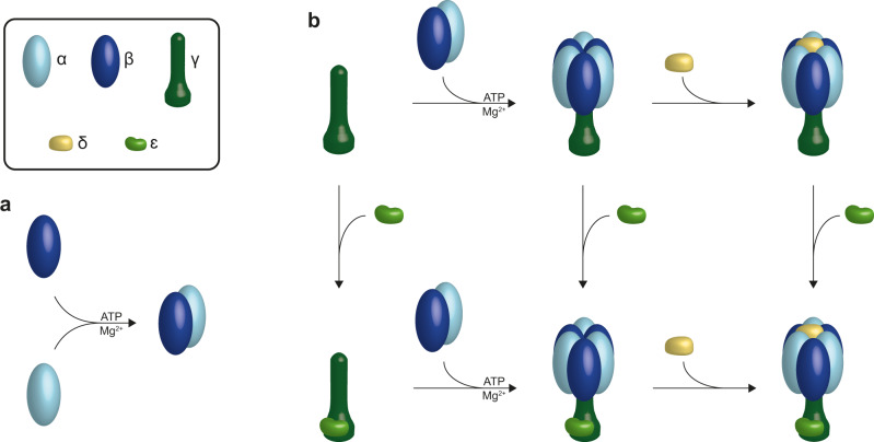
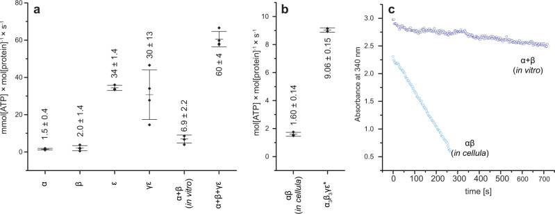
F-type ATP synthases are multiprotein complexes composed of two separate coupled motors (F1 and FO) generating adenosine triphosphate (ATP) as the universal major energy source in a variety of relevant biological processes in mitochondria, bacteria and chloroplasts. While the structure of many ATPases is solved today, the precise assembly pathway of F1FO-ATP synthases is still largely unclear. Here, we probe the assembly of the F1 complex from Acetobacterium woodii. Using laser induced liquid bead ion desorption (LILBID) mass spectrometry, we study the self-assembly of purified F1 subunits in different environments under non-denaturing conditions. We report assembly requirements and identify important assembly intermediates in vitro and in cellula. Our data provide evidence that nucleotide binding is crucial for in vitro F1 assembly, whereas ATP hydrolysis appears to be less critical. We correlate our results with activity measurements and propose a model for the assembly pathway of a functional F1 complex.


Conflict of interest statement

The authors declare no competing interests.

Figures

Fig. 1
Fig. 1. Dependence of ATP/Mg2+ in the assembly process of bacterial F-type ATP synthases.
a, b LILBID spectrum of in vitro α and β assembly: the α- (light blue) and β-subunit (dark blue) either without a or with b 2 mM ATP and 2 mM MgCl2. In presence of ATP/Mg2+ the mass spectrum shows the formation of a specific αβ heterodimer assembly, but no higher αβ oligomers. c High-performance Liquid Chromatography (HPLC) of in vitro α (light blue) and β (dark blue) assembly: the addition of 2 mM ATP and 2 mM MgCl2 to α and β lead to αβ heterodimer formation displayed by a shift from 12.27 min (light green area) to ~11.26 min (light red area). The elution peak at 13.25 min is owing to the high ATP concentration. d Model of bacterial F-type-ATPase: the soluble module F1 is organized by α- (light blue), β- (dark blue), γ- (dark green), δ- (yellow) and ε-subunit (light green). Source data underlying ac are provided as a Source Data file.
Fig. 2
Fig. 2. Summary of all subcomplexes obtained in vitro and in cellula.
a List of all subcomplexes observed with LILBID-MS after in vitro assembly of isolated subunits in the presence of 2 mM ATP and 2 mM MgCl2. b SDS-PAGE (NuPAGE 4–12% Bis-TRIS) of single purified subunits α, β, γε, and δ. Affinity purification of α with Strep-Tactin- and β, γε, and δ with Ni-chelating chromatography. N = 3 independent experiments. c List of in E. coli heterologously purified F1 and subcomplexes. All complexes were analyzed with LILBID-MS and SDS-PAGE (Supplementary Fig. 7). Source data underlying Fig. 2a are provided as a Source Data file.
Fig. 3
Fig. 3. ATP/Mg2+ dependence of the in vitro assembly process of the full F1 complex of Na+-F1FO- ATP synthases of A. woodii.
The α- (light blue), β-subunit (dark blue), γε-complex (dark and light green) were incubated at 4 °C for 1 h either without (a) or with (c) 2 mM ATP and 2 mM MgCl2. Then the δ-subunit (yellow) was added (b, d). a, b No specific assemblies into higher F1-subcomplexes could be detected without ATP/Mg2+. The appearance of the γ subunit alone is due to dissociation. (See as well Supplementary Fig. 8c). c, d In presence of ATP/Mg2+ the fully assembled F1 can be identified with additional F1 subcomplexes (e.g., α2β2γε, αβγε, αβγ) implying pairwise specific αβ heterodimer association. The δ-subunit can only bind when a complex containing the hexameric head domain α3β3 is already preformed. Source data are provided as a Source Data file.
Fig. 4
Fig. 4. Role of ATP hydrolysis vs. nucleotide-binding in the assembly process to F1 of bacterial F-type ATP synthases.
LILBID spectra of in vitro α, β, γε assembly: The α- (light blue), β-subunit (dark blue), γε-complex (dark and light green) were incubated with 2 mM MgCl2 and 2 mM of a nucleotide at 4 °C for 1 h. Complex formation is observed after incubation with ATP (a), AMP-PNP (b), ADP (c), and ATP-γ-S (d). Full α3β3γε assembly can be identified next to small amounts of other F1 subcomplexes (e.g., α2β2δγε α2β2γε, αβγε, αβγ), for ATP as well as the non-hydrolysable ATP analogs, apart from AMP-PMP, where the highest observed complex is α2β2γε. Source data are provided as a Source Data file.
Fig. 5
Fig. 5. Amino-acid sequence alignment of α- and β-subunit residues for E. coli and A. woodii.
a α-subunit residues. b β-subunit residues. Positions of positively charged amino acids that were mutated are marked in blue.
Fig. 6
Fig. 6. Dependence of ATP-binding of α, β and their mutants and the formation of αβ heterodimers.
a, b ATP-binding: deconvoluted nESI-MS spectra of ATP-binding of β[WT] (a) and β[K159Q] (b). Corresponding spectra for all α and β mutants are shown in Supplementary Fig. 4. c Summary of ATP-binding of β[K159Q], β[R186Q], β[R251Q], β[K159Q, R186Q, R251Q], α[WT], α[R363Q], and α[R363K] relative to β[WT]. Data are presented as mean values with error bars showing the SD, with individual data superimposed. dh HPLC measurements: in vitro αβ heterodimer assembly of α[WT] (light blue) with β[WT] (dark blue) and β mutants. i HPLC measurement: In vitro αβ heterodimer assembly of β[WT] (dark blue) with α[R363Q] (light blue). Data were collected in biological replicates (n = 3). Source data are provided as a Source Data file.
Fig. 7
Fig. 7. Role of charged residues in the catalytic site for the assembly process analyzed by LILBID-MS.
a In vitro assembly of α[WT] (light blue) with β[K159Q] mutant (dark blue) shows no significant formation of the αβ heterodimer, which we observe for the α[WT] and β[WT] (Fig. 1b). b The addition of the central stalk γε (dark and light green) does not lead to the formation of the desired α3β3γε complex. c In vitro assembled α[R363K] (light blue) and β[WT] (dark blue) revealed reduced αβ heterodimer assembly. d The biggest complex that is observed after addition of γε (dark and light green) is an α2β2γε subcomplex with charge states (−1 to −4). Source data are provided as a Source Data file.
Fig. 8
Fig. 8. Order of assembly steps into bacterial F1.
a As a first step α-subunits (light blue) and β-subunits (dark blue) form αβ heterodimers, which requires the presence of nucleotides and Mg2+. b As next step three of these heterodimer building blocks assemble pairwise to the central stalk γ (dark green). Only after the formation of the full hexameric head, the δ-subunit (yellow) is able to bind. The ε-subunit (light green) can join into the assembly process at all intermediate step en-route to the F1 complex.
Fig. 9
Fig. 9. ATP hydrolysis of isolated subunits and in vitro and in cellula assembled complexes.
a Isolated α, β, ε, γε and in vitro assembled αβ and α3β3γε complexes. b In cellula αβ and α3β3γε* complexes. Data are presented as mean values with error bars showing the SD, with individual data superimposed. c Spectroscopic comparison of the decrease in absorbance at 340 nm per time [s] in the enzyme-coupled ATPase activity assay of the in vitro assembled αβ from isolated α and β (5.5 µM each) relative to the in cellula produced αβ (0.5 µM protein complex) in 100 µL (100 mM TRIS, 100 mM maleic acid, 5 mM MgCl2, 3 mM phosphoenolpyruvate (PEP), 4 mM ATP, 0.5 mM NADH, 10 units L-lactate dehydrogenase (L-LDH), 10 units pyruvate kinase (PK), pH 7.5). In vitro data were collected in biological quadruplicates (n = 4) except ε with three biological replicates (n = 3) and in cellula data were collected in biological triplicates for αβ (n = 3) and technical duplicates for α3β3γε* (n = 2). Source data underlying Fig. 9a and b are provided as a Source Data file.
Fig. 10
Fig. 10. CIU fingerprints of the in cellula and in vitro assembled αβ heterodimer in the presence of ATP and Mg2+.
a In cellula assembled αβ heterodimer. b In vitro assembled αβ heterodimer. Exemplary spectra are shown in Supplementary Fig. 10. At low collision voltages the IM signal of the αβ heterodimer appears at 7595 Å2 for the in cellula and at 7630 Å2 for the in vitro assembled αβ heterodimer, respectively. At higher collision voltages the signal of the in vitro assembled αβ heterodimer unfolds, resulting in a signal at 7851 Å2. At 152.6 V 50% has reached the unfolded state. In contrast, increasing the collision voltage in the case of the in cellula formed αβ heterodimer leads to a signal appearing at 7802 Å2. 50% unfolding is never reached—the maximal unfolding of 47% is observed at 200 V. c The difference plot of both fingerprints illustrates the change regarding unfolding behavior between in vitro and in cellula assembled heterodimers. df CCS distribution for different collision voltages for in vitro (d) and in cellula (e) assembled heterodimers, and the difference plot (f). The overall feature of the in vitro αβ is clearly broader than for the in cellula αβ.

References

    1. Andries K. A Diarylquinoline drug active on the ATP synthase of Mycobacterium tuberculosis. Science. 2005;307:223–227. - PubMed
    1. De Jonge MR, Koymans LHM, Guillemont JEG, Koul A, Andries K. A computational model of the inhibition of Mycobacterium tuberculosis ATPase by a new drug candidate R207910. Proteins. 2007;67:971–980. - PubMed
    1. de Vries DD, van Engelen BGM, Gabreëls FJM, Ruitenbeek W, van Oost BA. A second missense mutation in the mitochondrial ATPase 6 gene in Leigh’s syndrome. Ann. Neurol. 1993;34:410–412. - PubMed
    1. Hong S, Pedersen PL. ATP synthase and the actions of inhibitors utilized to study its roles in human health, disease, and other scientific areas. Microbiol. Mol. Biol. Rev. 2008;72:590–641. - PMC - PubMed
    1. Cha MY, et al. Mitochondrial ATP synthase activity is impaired by suppressed O-GlcNAcylation in Alzheimer’s disease. Hum. Mol. Genet. 2015;24:6492–6504. - PMC - PubMed

Publication types

MeSH terms

Substances