. 2022 Mar 8;12(1):3729.
doi: 10.1038/s41598-022-07437-4.

Impact of silk hydrogel secondary structure on hydrogel formation, silk leaching and in vitro response

Affiliations

Impact of silk hydrogel secondary structure on hydrogel formation, silk leaching and in vitro response

Gemma Egan et al. Sci Rep. .

Abstract

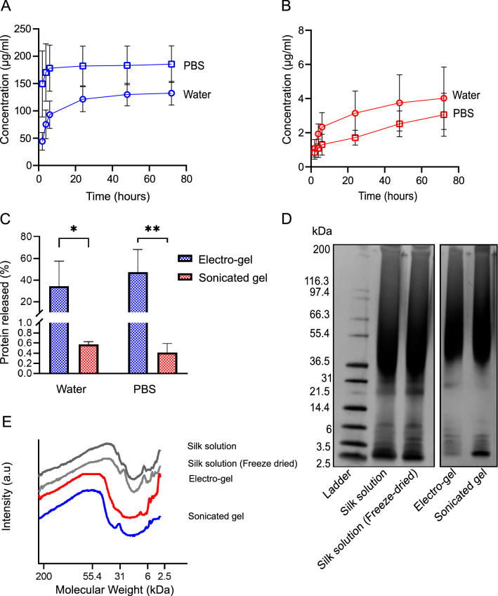
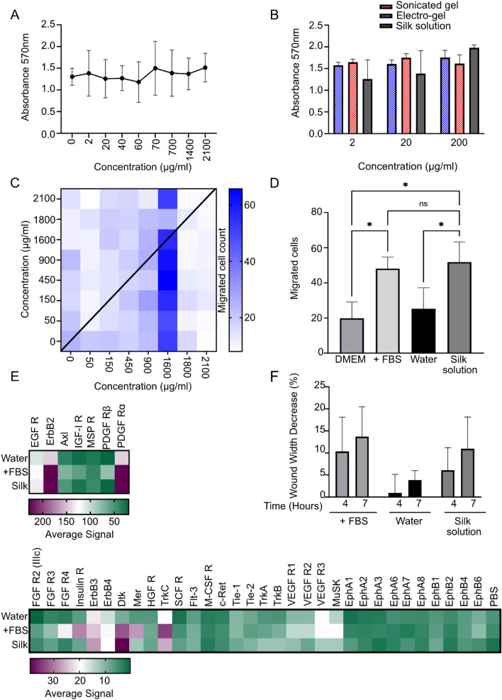
Silk can be processed into a broad spectrum of material formats and is explored for a wide range of medical applications, including hydrogels for wound care. The current paradigm is that solution-stable silk fibroin in the hydrogels is responsible for their therapeutic response in wound healing. Here, we generated physically cross-linked silk fibroin hydrogels with tuned secondary structure and examined their ability to influence their biological response by leaching silk fibroin. Significantly more silk fibroin leached from hydrogels with an amorphous silk fibroin structure than with a beta sheet-rich silk fibroin structure, although all hydrogels leached silk fibroin. The leached silk was biologically active, as it induced vitro chemokinesis and faster scratch assay wound healing by activating receptor tyrosine kinases. Overall, these effects are desirable for wound management and show the promise of silk fibroin and hydrogel leaching in the wider healthcare setting.


Conflict of interest statement

The authors declare no competing interests.

Figures

Figure 1
Figure 1
Production and characterisation of silk hydrogels. (A) Illustrative overview of the secondary structures found in silk solution and the changes seen with application of either ultrasonic waves or direct current, resulting in a sonicated hydrogel or an electro-gel, respectively (scale bar 0.25 cm). (B) Sol–gel efficiency of sonication or electro-gelation of silk fibroin solution (n = 5). (C) Solid content of the resultant electro-gel and sonicated gel (n = 5). (D) FTIR absorbance spectra of the amide I region of electro-gels and sonicated gels. Controls included were untreated air-dried silk film (UT) and the current treated solution remaining after removal of the electro-gel; this solution was air-dried into a film (CT). The third control was an ethanol treated silk film (EtOH) (n = 3). Leached samples in water of both electro-gel and sonicated gel after 72 h are included here. R is the correlation coefficient to freeze dried silk I. Lines at 1640 and 1621 indicate the amorphous and crystalline region, respectively (n = 8).
Figure 2
Figure 2
Protein release and characterisation from silk hydrogels measured by protein quantification and gel electrophoresis. (A) Electro-gel protein release in water and PBS over 72 h. (B) Sonicated hydrogel protein release in water and PBS over 72 h. (C) Total protein released after 72 h in water and PBS as a percentage of the starting quantity for electro-gels and sonicated gels (n = 4). (D) SDS PAGE of protein released from electro-gels and sonicated gels. Standards included are silk solution and freeze-dried silk solution reconstituted in water. (E) Densitometry analysis of SDS PAGE.
Figure 3
Figure 3
In vitro studies with silk fibroin. NIH 3T3 mouse fibroblasts were used throughout. (A) Cell proliferation of NIH3T3 fibroblasts incubated with silk solution for 72 h and cell viability measured with an MTT assay (n = 3). (B) Cell proliferation of NIH3T3 fibroblasts incubated with leached samples of silk from electro-gels, sonicated gels or silk solution after 72 h (n = 3). (C) Checkerboard migration assay. Cell migration across a permeable membrane after exposure to silk solution for 3.5 h (n = 4). The X and Y axes depict silk concentrations above and below the filter, respectively. Below the filter is the receiver chamber. (D) Cell migration at 1600 µg/mL silk solution and controls with the presence of FBS as a positive control and water as the negative control (n = 4). (E) Phosphorylation array in the presence of silk solution, water or FBS. (F) Wound closure assay width over 4 and 7 h in the presence of silk solution, water or FBS (n = 3).

References

    1. Carter MJ, Tingley-Kelley K, Warriner RA., 3rd Silver treatments and silver-impregnated dressings for the healing of leg wounds and ulcers: A systematic review and meta-analysis. J. Am. Acad. Dermatol. 2010;63:668–679. doi: 10.1016/j.jaad.2009.09.007. - DOI - PubMed
    1. Amsler F, Willenberg T, Blattler W. In search of optimal compression therapy for venous leg ulcers: A meta-analysis of studies comparing diverse [corrected] bandages with specifically designed stockings. J. Vasc. Surg. 2009;50:668–674. doi: 10.1016/j.jvs.2009.05.018. - DOI - PubMed
    1. Cullum N, Liu Z. Therapeutic ultrasound for venous leg ulcers. Cochrane Database Syst. Rev. 2017;5:CD001180. doi: 10.1002/14651858.CD001180.pub4. - DOI - PMC - PubMed
    1. Mohd Zubir MZ, Holloway S, Mohd Noor N. Maggot therapy in wound healing: A systematic review. Int. J. Environ. Res. Public Health. 2020;17:6103. doi: 10.3390/ijerph17176103. - DOI - PMC - PubMed
    1. Greaves NS, Benatar B, Baguneid M, Bayat A. Single-stage application of a novel decellularized dermis for treatment-resistant lower limb ulcers: Positive outcomes assessed by SIAscopy, laser perfusion, and 3D imaging, with sequential timed histological analysis. Wound Repair Regen. 2013;21:813–822. doi: 10.1111/wrr.12113. - DOI - PubMed

Publication types