. 2022 Mar 8;12(1):3770.
doi: 10.1038/s41598-022-07630-5.

Stability, dissolution, and cytotoxicity of NaYF4-upconversion nanoparticles with different coatings

Affiliations

Stability, dissolution, and cytotoxicity of NaYF4-upconversion nanoparticles with different coatings

Verónica Bastos et al. Sci Rep. .

Abstract

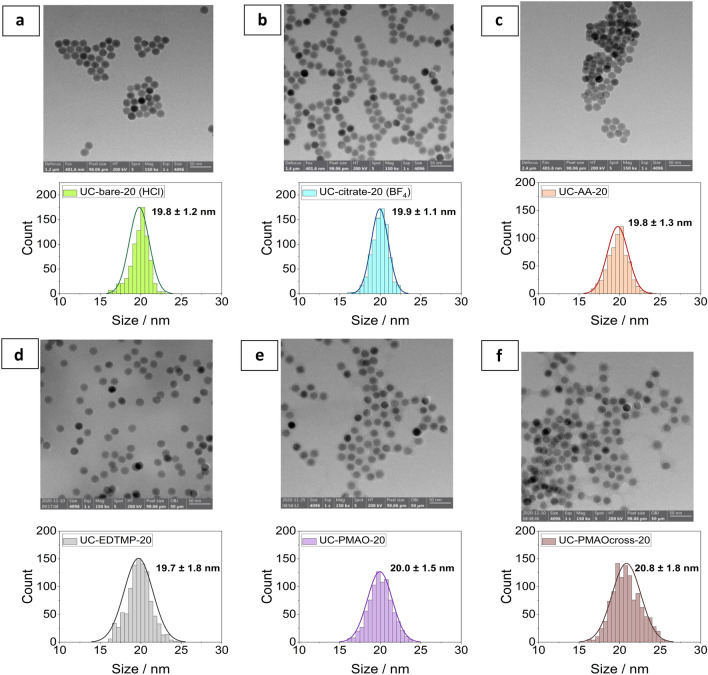
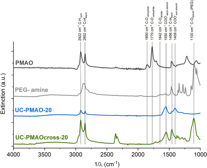
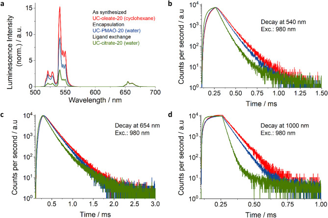
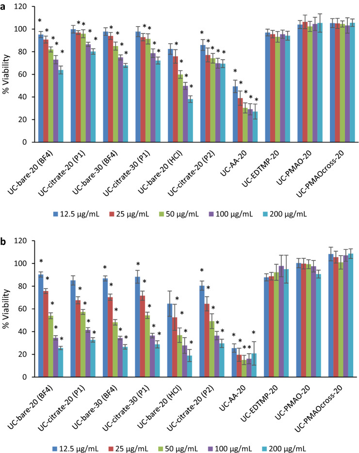
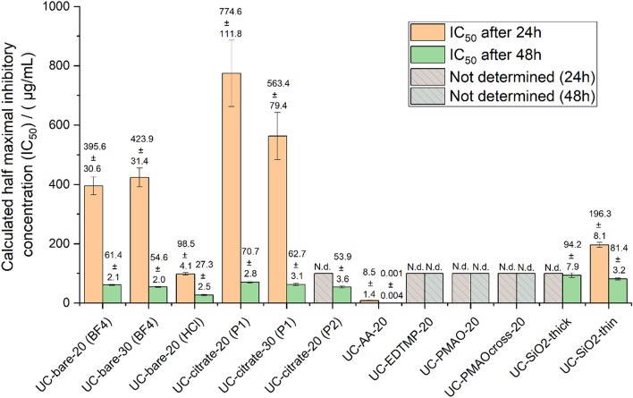
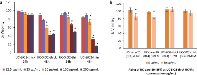
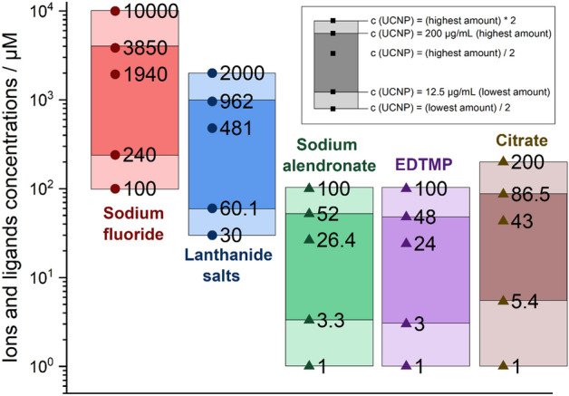
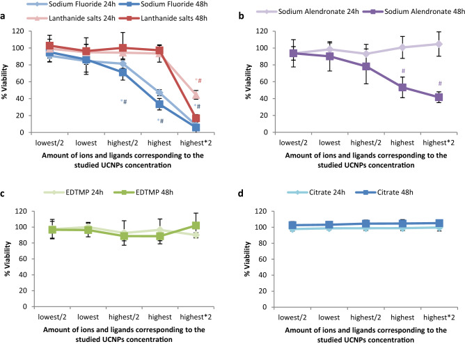
Upconversion nanoparticles (UCNPs) have attracted considerable attention owing to their unique photophysical properties. Their utilization in biomedical applications depends on the understanding of their transformations under physiological conditions and their potential toxicity. In this study, NaYF4:Yb,Er UCNPs, widely used for luminescence and photophysical studies, were modified with a set of four different coordinatively bound surface ligands, i.e., citrate, alendronate (AA), ethylendiamine tetra(methylene phosphonate) (EDTMP), and poly(maleic anhydride-alt-1-octadecene) (PMAO), as well as silica coatings with two different thicknesses. Subsequently, the aging-induced release of fluoride ions in water and cell culture media and their cytotoxic profile to human keratinocytes were assessed in parallel to the cytotoxic evaluation of the ligands, sodium fluoride and the lanthanide ions. The cytotoxicity studies of UCNPs with different surface modifications demonstrated the good biocompatibility of EDTMP-UCNPs and PMAO-UCNPs, which is in line with the low amount of fluoride ions released from these samples. An efficient prevention of UCNP dissolution and release of cytotoxic ions, as well as low cytotoxicity was also observed for UCNPs with a sufficiently thick silica shell. Overall, our results provide new insights into the understanding of the contribution of surface chemistry to the stability, dissolution behavior, and cytotoxicity of UCNPs. Altogether, the results obtained are highly important for future applications of UCNPs in the life sciences and bioimaging studies.


Conflict of interest statement

The authors declare no competing interests.

Figures

Figure 1
Figure 1
Transmission electron microscopy (TEM) images of UCNP with different surface coatings. (a) UC-bare-20 (HCl); (b) UC-citrate-20 (BF4); (c) UC-AA-20; (d) UC-EDTMP-20; (e) UC-PMAO-20; (f) UC-PMAOcross-20.
Figure 2
Figure 2
ATR-FTIR spectra of UCNPs with amphiphilic coatings and the corresponding ligands.
Figure 3
Figure 3
Luminescence properties of the differently coated hydrophilic UCNPs modified via encapsulation (blue) and ligand exchange (green) compared to the as-synthesized hydrophobic UCNPs. (a) Photoluminescence spectra measured at an excitation power density (P) of 22 W/cm2 using a 980 nm laser diode; the spectra were normalized at 654 nm. (b) Lifetime of the Er3+ upconversion luminescence (UCL) excited at 980 nm (P of 67 W/cm2; pulse width 40 µs) and recorded at 540 nm; (c) Lifetime of the Er3+ UCL excited at 980 nm (P of 128 W/cm2; pulse width 40 µs) recorded at 655 nm; and d) Lifetime of the downshifted luminescence (DSL) of Yb3+ excited at 980 nm (P of 128 W/cm2; pulse width 40 µs) and recorded at 1000 nm.
Figure 4
Figure 4
Viability of HaCaT cells after exposure to UCNPs with different surface coatings for (a) 24 h and (b) 48 h: UC-bare-20 (BF4); UC-citrate-20 (P1); UC-bare-30 (BF4); UC-citrate-30 (P1); UC-bare-20 (HCl); UC-citrate-20 (P2); UC-AA-20; UC-EDTMP-20; UC-PMAO-20; UC-PMAOcross-20. The results are reported as mean ± standard deviation (SD) of 4 technical replicates in each of the 3 independent experiments for 24 h and 48 h. * indicates significant statistically differences compared to the control (p < 0.05).
Figure 5
Figure 5
Calculated half maximal inhibitory concentration (IC50) at 24 h and 48 h exposure with UCNPs of different surface modification. N.d.: not determined.
Figure 6
Figure 6
Viability of HaCaT cells exposed to (a) UC-SiO2-thick and UC-SiO2-thin for 24 h and 48 h; and (b) UC-bare-20 (BF4) and UC-SiO2-thick after 72 h of aging in di-H2O and DMEM. The results are reported as mean ± standard deviation (SD) of 4 technical replicates in each of the 3 independent experiments for 24 h and 48 h. The differences were considered statistically significant from p < 0.05: * indicates significant differences compared to the control.
Figure 7
Figure 7
Concentration range of ions and ligands (µM) used for viability tests based on calculations of their total amount present in the lowest (12.5 µg/mL) and highest (200 µg/mL) concentration of UCNPs for the studied particles, assuming their complete release.
Figure 8
Figure 8
Thermogravimetric analysis (TGA) curves of 21.5 nm-sized NaYF4:Yb,Er particles with citrate coating (procedure 2).
Figure 9
Figure 9
Viability of HaCaT cells after exposure to the UCNP constituting ions and ligands at concentrations matching the concentration range of released upon exposure of the UCNPs; the lowest amount (amount present in the 12.5 µg/mL of UCNPs), half of the lowest amount, half of the highest amount, highest amount (amount present in the 200 µg/mL of UCNPs), and twice of the highest amount. a) Sodium fluoride and lanthanide salts; b) Sodium alendronate; c) EDTMP; d) Citrate. The results are reported as mean ± standard deviation (SD) of 4 replicates from each of the 3 independent experiments for 24 h and 48 h. The differences were considered statistically significant from p < 0.05: * indicates significant differences compared to the control after 24 h, # after 48 h.

References

    1. Ao Y, et al. An upconversion nanoparticle enables near infrared-optogenetic manipulation of the Caenorhabditis elegans motor circuit. ACS Nano. 2019;13:3373–3386. doi: 10.1021/acsnano.8b09270. - DOI - PubMed
    1. Mahata MK, De R, Lee KT. Near-infrared-triggered upconverting nanoparticles for biomedicine applications. Biomedicines. 2021;9:756. doi: 10.3390/biomedicines9070756. - DOI - PMC - PubMed
    1. Jafari M, Rezvanpour A. Upconversion nano-particles from synthesis to cancer treatment: A review. Adv. Powder Technol. 2019;30:1731–1753. doi: 10.1016/j.apt.2019.05.027. - DOI
    1. Han R, et al. Fabrication of core/shell/shell structure nanoparticle with anticancer drug and dual-photosensitizer co-loading for synergistic chemotherapy and photodynamic therapy. Microporous Mesoporous Mater. 2020;297:110049. doi: 10.1016/j.micromeso.2020.110049. - DOI
    1. Liang G, et al. Recent progress in the development of upconversion nanomaterials in bioimaging and disease treatment. J. Nanobiotechnol. 2020;18:1–22. doi: 10.1186/s12951-019-0560-5. - DOI - PMC - PubMed

Publication types