

ACE2 protein expression in lung tissues of severe COVID-19 infection
- PMID: 35260724
- PMCID: PMC8902283
- DOI: 10.1038/s41598-022-07918-6
ACE2 protein expression in lung tissues of severe COVID-19 infection
Abstract
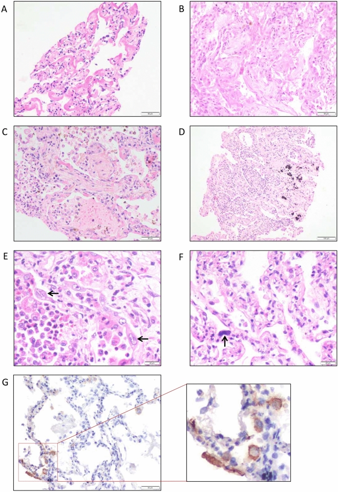
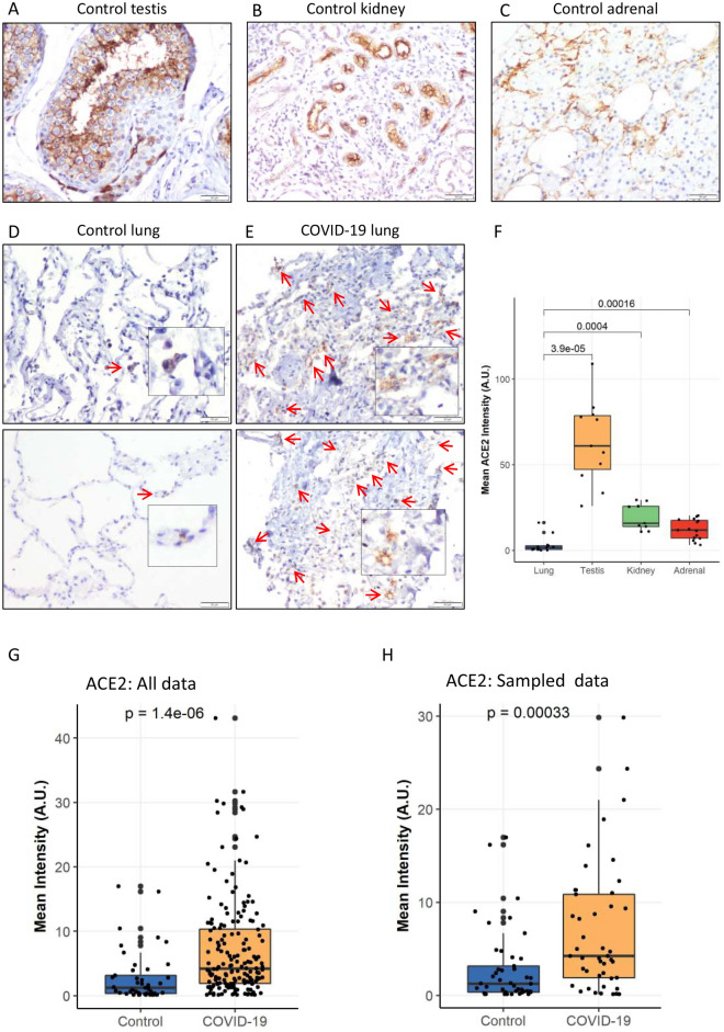
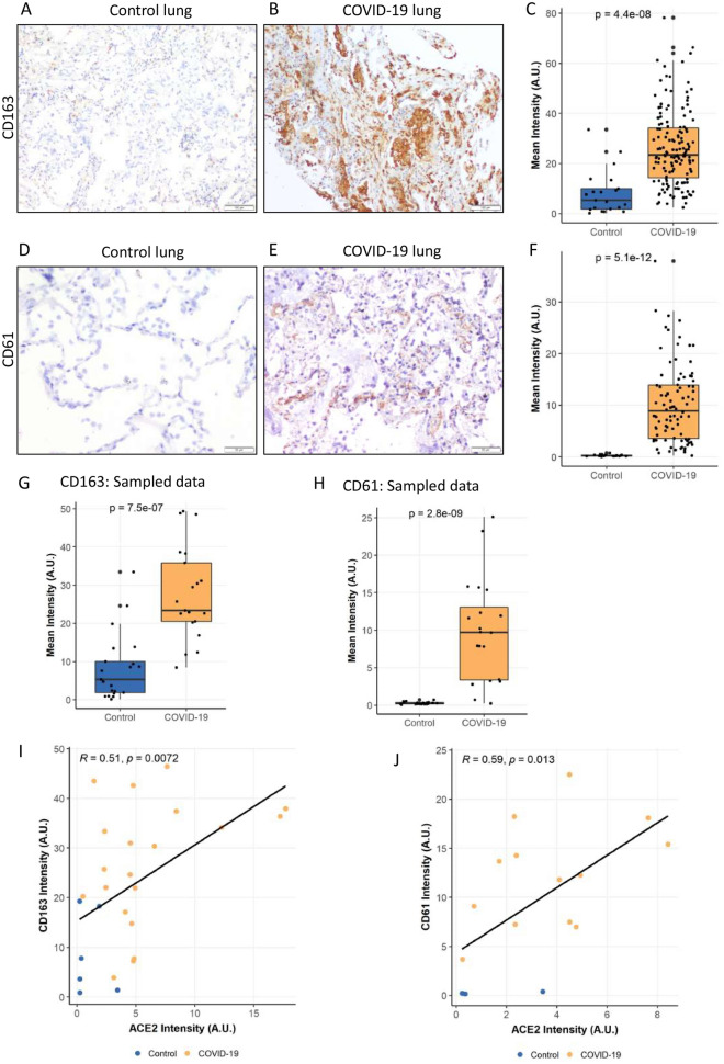
Angiotensin-converting enzyme 2 (ACE2) is a key host protein by which severe acute respiratory syndrome coronavirus-2 (SARS-CoV-2) enters and multiplies within cells. The level of ACE2 expression in the lung is hypothesised to correlate with an increased risk of severe infection and complications in COrona VIrus Disease 2019 (COVID-19). To test this hypothesis, we compared the protein expression status of ACE2 by immunohistochemistry (IHC) in post-mortem lung samples of patients who died of severe COVID-19 and lung samples obtained from non-COVID-19 patients for other indications. IHC for CD61 and CD163 was performed for the assessment of platelet-rich microthrombi and macrophages, respectively. IHC for SARS-CoV-2 viral antigen was also performed. In a total of 55, 44 COVID-19 post-mortem lung samples were tested for ACE2, 36 for CD163, and 26 for CD61, compared to 15 non-covid 19 control lung sections. Quantification of immunostaining, random sampling, and correlation analysis were used to substantiate the morphologic findings. Our results show that ACE2 protein expression was significantly higher in COVID-19 post-mortem lung tissues than in controls, regardless of sample size. Histomorphology in COVID-19 lungs showed diffuse alveolar damage (DAD), acute bronchopneumonia, and acute lung injury with SARS-CoV-2 viral protein detected in a subset of cases. ACE2 expression levels were positively correlated with increased expression levels of CD61 and CD163. In conclusion, our results show significantly higher ACE2 protein expression in severe COVID-19 disease, correlating with increased macrophage infiltration and microthrombi, suggesting a pathobiological role in disease severity.
© 2022. The Author(s).
Conflict of interest statement
The authors declare no competing interests.
Figures
Similar articles
-
Overexpression of the SARS-CoV-2 receptor ACE2 is induced by cigarette smoke in bronchial and alveolar epithelia.J Pathol. 2021 Jan;253(1):17-30. doi: 10.1002/path.5555. Epub 2020 Oct 27. J Pathol. 2021. PMID: 32991738 Free PMC article.
-
Hepatic angiotensin-converting enzyme 2 expression in metabolic dysfunction-associated steatotic liver disease and in patients with fatal COVID-19.World J Gastroenterol. 2024 Aug 21;30(31):3705-3716. doi: 10.3748/wjg.v30.i31.3705. World J Gastroenterol. 2024. PMID: 39192998 Free PMC article.
-
Dysbalance of ACE2 levels - a possible cause for severe COVID-19 outcome in COPD.J Pathol Clin Res. 2021 Sep;7(5):446-458. doi: 10.1002/cjp2.224. Epub 2021 May 12. J Pathol Clin Res. 2021. PMID: 33978304 Free PMC article.
-
Effects of SARS-CoV-2 on Cardiovascular System: The Dual Role of Angiotensin-Converting Enzyme 2 (ACE2) as the Virus Receptor and Homeostasis Regulator-Review.Int J Mol Sci. 2021 Apr 26;22(9):4526. doi: 10.3390/ijms22094526. Int J Mol Sci. 2021. PMID: 33926110 Free PMC article. Review.
-
The expression of hACE2 receptor protein and its involvement in SARS-CoV-2 entry, pathogenesis, and its application as potential therapeutic target.Tumour Biol. 2021;43(1):177-196. doi: 10.3233/TUB-200084. Tumour Biol. 2021. PMID: 34420993 Review.
Cited by
-
Persisting Shadows: Unraveling the Impact of Long COVID-19 on Respiratory, Cardiovascular, and Nervous Systems.Infect Dis Rep. 2023 Dec 15;15(6):806-830. doi: 10.3390/idr15060072. Infect Dis Rep. 2023. PMID: 38131885 Free PMC article. Review.
-
Vascular Implications of COVID-19: Role of Radiological Imaging, Artificial Intelligence, and Tissue Characterization: A Special Report.J Cardiovasc Dev Dis. 2022 Aug 15;9(8):268. doi: 10.3390/jcdd9080268. J Cardiovasc Dev Dis. 2022. PMID: 36005433 Free PMC article. Review.
-
HDL-Related Parameters and COVID-19 Mortality: The Importance of HDL Function.Antioxidants (Basel). 2023 Nov 16;12(11):2009. doi: 10.3390/antiox12112009. Antioxidants (Basel). 2023. PMID: 38001862 Free PMC article.
-
Mucosal Immunity against SARS-CoV-2 in the Respiratory Tract.Pathogens. 2024 Jan 26;13(2):113. doi: 10.3390/pathogens13020113. Pathogens. 2024. PMID: 38392851 Free PMC article. Review.
-
Dramatic Decrease of Vitamin K2 Subtype Menaquinone-7 in COVID-19 Patients.Antioxidants (Basel). 2022 Jun 24;11(7):1235. doi: 10.3390/antiox11071235. Antioxidants (Basel). 2022. PMID: 35883726 Free PMC article.
References
Publication types
MeSH terms
Substances
Grants and funding
- A-COVID 40/This work is supported by intramural funding from the research section of the All India Institute of Medical Sciences (AIIMS), New Delhi, India.
- A-COVID 40/This work is supported by intramural funding from the research section of the All India Institute of Medical Sciences (AIIMS), New Delhi, India.
- A-COVID 40/This work is supported by intramural funding from the research section of the All India Institute of Medical Sciences (AIIMS), New Delhi, India.
- A-COVID 40/This work is supported by intramural funding from the research section of the All India Institute of Medical Sciences (AIIMS), New Delhi, India.
- A-COVID 40/This work is supported by intramural funding from the research section of the All India Institute of Medical Sciences (AIIMS), New Delhi, India.
- A-COVID 40/This work is supported by intramural funding from the research section of the All India Institute of Medical Sciences (AIIMS), New Delhi, India.
- A-COVID 40/This work is supported by intramural funding from the research section of the All India Institute of Medical Sciences (AIIMS), New Delhi, India.
- A-COVID 40/This work is supported by intramural funding from the research section of the All India Institute of Medical Sciences (AIIMS), New Delhi, India.
- A-COVID 40/This work is supported by intramural funding from the research section of the All India Institute of Medical Sciences (AIIMS), New Delhi, India.
- A-COVID 40/This work is supported by intramural funding from the research section of the All India Institute of Medical Sciences (AIIMS), New Delhi, India.
- A-COVID 40/This work is supported by intramural funding from the research section of the All India Institute of Medical Sciences (AIIMS), New Delhi, India.
- A-COVID 40/This work is supported by intramural funding from the research section of the All India Institute of Medical Sciences (AIIMS), New Delhi, India.
- A-COVID 40/This work is supported by intramural funding from the research section of the All India Institute of Medical Sciences (AIIMS), New Delhi, India.
- A-COVID 40/This work is supported by intramural funding from the research section of the All India Institute of Medical Sciences (AIIMS), New Delhi, India.
- A-COVID 40/This work is supported by intramural funding from the research section of the All India Institute of Medical Sciences (AIIMS), New Delhi, India.
- A-COVID 40/This work is supported by intramural funding from the research section of the All India Institute of Medical Sciences (AIIMS), New Delhi, India.
- A-COVID 40/This work is supported by intramural funding from the research section of the All India Institute of Medical Sciences (AIIMS), New Delhi, India.
- A-COVID 40/This work is supported by intramural funding from the research section of the All India Institute of Medical Sciences (AIIMS), New Delhi, India.
LinkOut - more resources
Full Text Sources
Medical
Research Materials
Miscellaneous
