

PCDHB15 as a potential tumor suppressor and epigenetic biomarker for breast cancer
- PMID: 35261631
- PMCID: PMC8855166
- DOI: 10.3892/ol.2022.13237
PCDHB15 as a potential tumor suppressor and epigenetic biomarker for breast cancer
Abstract
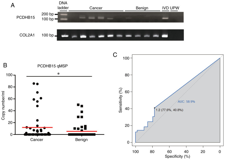
Breast cancer is among the most frequently diagnosed cancer types and the leading cause of cancer-related death in women. The mortality rate of patients with breast cancer is currently increasing, perhaps due to a lack of early screening tools. In the present study, using The Cancer Genome Atlas (TCGA) breast cancer dataset (n=883), it was determined that methylation of the protocadherin β15 (PCDHB15) promoter was higher in breast cancer samples than that in normal tissues. A negative association between promoter methylation and expression of PCDHB15 was observed in the TCGA dataset and breast cancer cell lines. In TCGA cohort, lower PCDHB15 expression was associated with shorter relapse-free survival times. Treatment with the DNA methyltransferase inhibitor restored PCDHB15 expression in a breast cancer cell line; however, overexpression of PCDHB15 was shown to suppress colony formation. PCDHB15 methylation detected in circulating cell-free DNA (cfDNA) isolated from serum samples was higher in patients with breast cancer (40.8%) compared with that in patients with benign tumors (22.4%). PCDHB15 methylation was not correlated with any clinical parameters. Taken together, PCDHB15 is a potential tumor suppressor in cases of breast cancer, which can be epigenetically silenced via promoter methylation. PCDHB15 methylation using cfDNA is a novel minimally invasive epigenetic biomarker for the diagnosis and prognosis of breast cancer.
Keywords: DNA methylation; biomarker; breast cancer; cell-free DNA; protocadherin β15.
Copyright: © Chiang et al.
Conflict of interest statement
The authors declare that they have no competing interests.
Figures


Similar articles
-
Epigenetically regulated PCDHB15 impairs aggressiveness of metastatic melanoma cells.Clin Epigenetics. 2022 Nov 28;14(1):156. doi: 10.1186/s13148-022-01364-x. Clin Epigenetics. 2022. PMID: 36443814 Free PMC article.
-
A genome-wide cell-free DNA methylation analysis identifies an episignature associated with metastatic luminal B breast cancer.Front Cell Dev Biol. 2022 Oct 25;10:1016955. doi: 10.3389/fcell.2022.1016955. eCollection 2022. Front Cell Dev Biol. 2022. PMID: 36393855 Free PMC article.
-
Circulating cell-free DNA-based epigenetic assay can detect early breast cancer.Breast Cancer Res. 2016 Dec 19;18(1):129. doi: 10.1186/s13058-016-0788-z. Breast Cancer Res. 2016. PMID: 27993161 Free PMC article.
-
A Systematic Analysis of the Relationship of CDH13 Promoter Methylation and Breast Cancer Risk and Prognosis.PLoS One. 2016 May 6;11(5):e0149185. doi: 10.1371/journal.pone.0149185. eCollection 2016. PLoS One. 2016. PMID: 27153114 Free PMC article. Review.
-
Early Epigenetic Markers for Precision Medicine.Methods Mol Biol. 2018;1856:3-17. doi: 10.1007/978-1-4939-8751-1_1. Methods Mol Biol. 2018. PMID: 30178243 Review.
Cited by
-
Differential methylation pattern in pubertal girls associated with biochemical premature adrenarche.Epigenetics. 2023 Dec;18(1):2200366. doi: 10.1080/15592294.2023.2200366. Epigenetics. 2023. PMID: 37053179 Free PMC article.
-
The Dysregulation of SOX Family Correlates with DNA Methylation and Immune Microenvironment Characteristics to Predict Prognosis in Hepatocellular Carcinoma.Dis Markers. 2022 Apr 13;2022:2676114. doi: 10.1155/2022/2676114. eCollection 2022. Dis Markers. 2022. PMID: 35465267 Free PMC article.
-
Liquid Biopsy-Based DNA Methylation Biomarkers for Precision Medicine in Breast Cancer.Expert Rev Mol Med. 2025 Jun 17;27:e20. doi: 10.1017/erm.2025.10008. Expert Rev Mol Med. 2025. PMID: 40524349 Free PMC article. Review.
-
Epigenetically regulated PCDHB15 impairs aggressiveness of metastatic melanoma cells.Clin Epigenetics. 2022 Nov 28;14(1):156. doi: 10.1186/s13148-022-01364-x. Clin Epigenetics. 2022. PMID: 36443814 Free PMC article.
-
Elucidating Novel Targets for Ovarian Cancer Antibody-Drug Conjugate Development: Integrating In Silico Prediction and Surface Plasmon Resonance to Identify Targets with Enhanced Antibody Internalization Capacity.Antibodies (Basel). 2023 Oct 16;12(4):65. doi: 10.3390/antib12040065. Antibodies (Basel). 2023. PMID: 37873862 Free PMC article.
References
-
- Chen YP, Lu YW, Yang CC. Breast cancer trend in Taiwan. MOJ Womens Health. 2017;6:153.
LinkOut - more resources
Full Text Sources