Ultrastructural, Histochemical, Cytological Study of Retina of Aborted Fetus of Various Weeks of Gestation - an Anatomical Perspective with Implications on Patients with Retinitis Pigmentosa
- PMID: 35261668
- PMCID: PMC8897797
- DOI: 10.26574/maedica.2021.16.4.656
Ultrastructural, Histochemical, Cytological Study of Retina of Aborted Fetus of Various Weeks of Gestation - an Anatomical Perspective with Implications on Patients with Retinitis Pigmentosa
Abstract
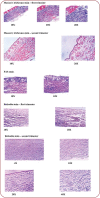
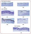
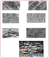
Aim: To study the ultrastructural, histochemical, cytological features of retina in aborted fetuses of different gestational age and its probable implication in the disease process of retinitis pigmentosa. Methodology: This is a prospective randomized cross sectional study that has been carried out in AIIMS Bhubaneswar from June 2017 to May 2019, jointly by the Department of Anatomy and Obstetrics and Gynaecology, AIIMS, Bhubaneswar, India, after proper institutional approval. Three fetuses from each trimester were taken into the present study; their retina was collected and subsequently sent for cytological, immunohistochemical and ultrastructural explorations. Detailed information from all explorations were collected and properly documented. Results:Fetuses whose retinas that have been shown to contain very few to no rod cells and short-sized cone cells might tend to develop retinitis pigmentosa after birth. Moreover, those cone cells have been shown to contain melanolysosome, phagosomes, autophagic vacuoles, and membranous whorls.
Figures
Similar articles
-
Cytological, Histochemical, and Ultrastructural Study of the Human Fetal Spleen of Various Gestational Age With Future Implications in Splenic Transplantation: An Anatomical Perspective.Cureus. 2021 Oct 19;13(10):e18911. doi: 10.7759/cureus.18911. eCollection 2021 Oct. Cureus. 2021. PMID: 34820226 Free PMC article.
-
Histopathology of the human retina in retinitis pigmentosa.Prog Retin Eye Res. 1998 Apr;17(2):175-205. doi: 10.1016/s1350-9462(97)00012-8. Prog Retin Eye Res. 1998. PMID: 9695792 Review.
-
Disease expression in X-linked retinitis pigmentosa caused by a putative null mutation in the RPGR gene.Invest Ophthalmol Vis Sci. 1997 Sep;38(10):1983-97. Invest Ophthalmol Vis Sci. 1997. PMID: 9331262
-
Autosomal dominant retinitis pigmentosa caused by the threonine-17-methionine rhodopsin mutation: retinal histopathology and immunocytochemistry.Exp Eye Res. 1994 Apr;58(4):397-408. doi: 10.1006/exer.1994.1032. Exp Eye Res. 1994. PMID: 7925677
-
Wide-Field Fundus Autofluorescence for Retinitis Pigmentosa and Cone/Cone-Rod Dystrophy.Adv Exp Med Biol. 2016;854:307-13. doi: 10.1007/978-3-319-17121-0_41. Adv Exp Med Biol. 2016. PMID: 26427426 Review.
References
-
- Curcio CA, Sloan KR, Hendrickson AE, Kalina RE. Individual variability in topography of human retinal photoreceptors. Soc Neurosci Abstr. 1986;12:636.
-
- Curcio CE, Sloan KR, Kalina RE, Hendrickson A. Human photoreceptor topography. J Comp Neurol. 1990;292:497–523. - PubMed
-
- Curcio CA, Hendrickson A. Organization and development of the primate photoreceptor mosaic. Prog Retin Res. 1991;10:90–120.
-
- Provis JM, Diaz CM, Dreher B. Ontogeny of the primate fovea: a central issue in retinal development. Prog Neurobiol. 1998;54:549–580. - PubMed
-
- Osterberg GA. Topography of the layer of rods and cones in the human retina. Acta Ophthalmologica. 1935;13:1–97.
Publication types
LinkOut - more resources
Full Text Sources


