

Substantial Atrophy of the Psoas Muscle as Late Sequela of L2 Osteoporotic Fracture: a Case Report
- PMID: 35261681
- PMCID: PMC8897803
- DOI: 10.26574/maedica.2020.16.4.738
Substantial Atrophy of the Psoas Muscle as Late Sequela of L2 Osteoporotic Fracture: a Case Report
Abstract
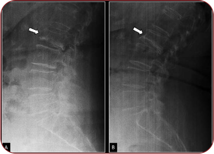
Osteoporotic vertebral fractures (OVFs) are considered benign and heal after 8-12 weeks. Nevertheless, up to one third of these patients will have persistent back pain, which may be complicated with neurologic deficit or paraplegia A unique unusual case of delayed onset of neurological complication of an osteoporotic vertebral fracture (OVF) in an elderly patient is reported. The patient presented with paraparesis due to isolated substantial atrophy of the psoas muscle 12 months after the initial fracture. The patient was investigated with imaging and nerve contacted studies. We suggest that psoas muscle atrophy can be determinant clinical sign to diagnose neurological compromise resulting from OVF, even if there is no other clinical indicators of spinal pathology.
Figures

Similar articles
-
Symptomatic relevance of intravertebral cleft in patients with osteoporotic vertebral fracture.J Neurosurg Spine. 2010 Aug;13(2):267-75. doi: 10.3171/2010.3.SPINE09364. J Neurosurg Spine. 2010. PMID: 20672965
-
Percutaneous kyphoplasty for the treatment of osteoporotic thoracolumbar fractures with neurological deficit: radicular pain can mimic disc herniation.Int J Clin Exp Med. 2014 Aug 15;7(8):2360-4. eCollection 2014. Int J Clin Exp Med. 2014. PMID: 25232437 Free PMC article.
-
The variability of vertebral body volume and pain associated with osteoporotic vertebral fractures: conservative treatment versus percutaneous transpedicular vertebroplasty.Int Orthop. 2017 May;41(5):963-968. doi: 10.1007/s00264-017-3409-2. Epub 2017 Feb 4. Int Orthop. 2017. PMID: 28161853
-
[Back pain and neurological deficits in osteoporotic spinal fractures].Hokkaido Igaku Zasshi. 1997 Jul;72(4):381-7. Hokkaido Igaku Zasshi. 1997. PMID: 9266250 Review. Japanese.
-
Radiographic diagnosis of osteoporotic vertebral fractures. An updated review.Med Clin (Barc). 2022 Feb 11;158(3):125-132. doi: 10.1016/j.medcli.2021.06.019. Epub 2021 Aug 13. Med Clin (Barc). 2022. PMID: 34392986 Review. English, Spanish.
References
-
- Alpantaki K, Dohm M, Korovessis P, Hadjipavlou AG. Surgical options for osteoporotic vertebral compression fractures complicated with spinal deformity and neurologic deficit. Injury. 2018;49:261–271. - PubMed
-
- Heggeness MH. Spine fracture with neurological deficit in osteoporosis. Osteoporos Int. 1993;3:215–221. - PubMed
-
- Baba H, Maezawa Y, Kamitani K, et al. Osteoporotic vertebral collapse with late neurological complications. Paraplegia. 1995;33:281–289. - PubMed
-
- Korovessis P, Maraziotis T, Piperos G, Spyropoulos P. Spontaneous burst fracture of the thoracolumbar spine in osteoporosis associated with neurological impairment: a report of seven cases and review of the literature. Eur Spine J. 1994;3:286–288. - PubMed
Publication types
LinkOut - more resources
Full Text Sources