Review
. 2022 Feb 15;12(2):861-872.
eCollection 2022.

Glycolysis in the progression of pancreatic cancer

Affiliations
Review

Glycolysis in the progression of pancreatic cancer

Xinyao Chang et al. Am J Cancer Res. .

Abstract

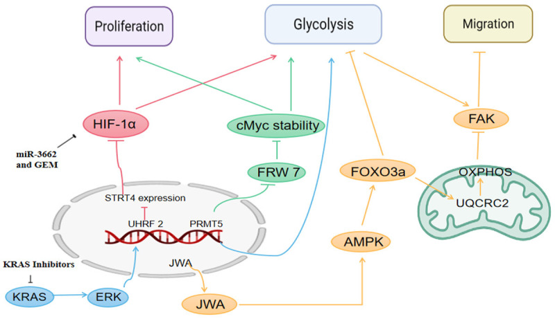
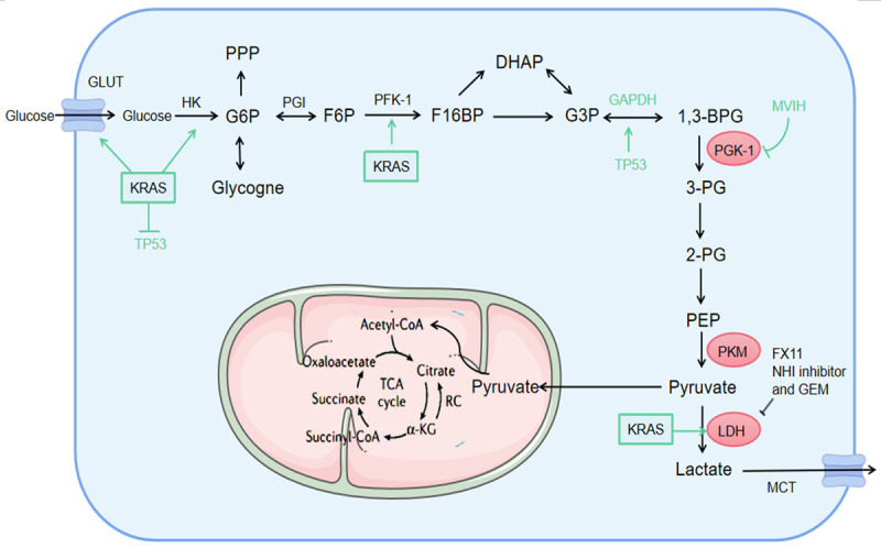
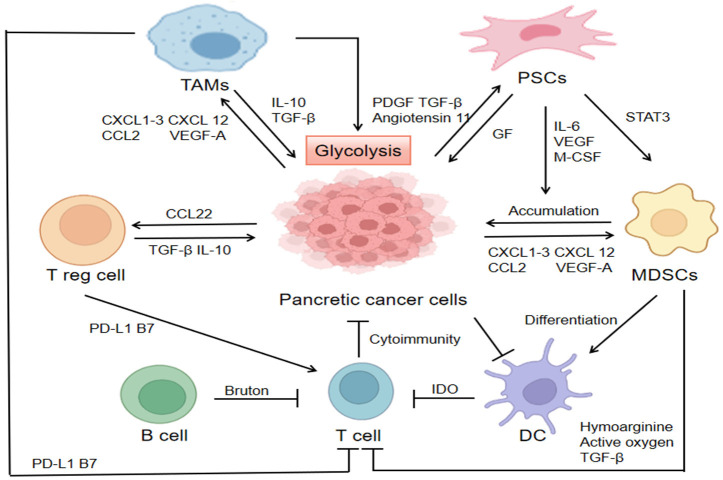
Metabolic reprogramming, as a key hallmark of cancers, leads to the malignant behavior of pancreatic cancer, which is closely related to tumor development and progression, as well as the supportive tumor microenvironments. Although cells produce adenosine triphosphate (ATP) from glucose by glycolysis when lacking oxygen, pancreatic cancer cells elicit metabolic conversion from oxide phosphorylation to glycolysis, which is well-known as "Warburg effect". Glycolysis is critical for cancer cells to maintain their robust biosynthesis and energy requirement, and it could promote tumor initiation, invasion, angiogenesis, and metastasis to distant organs. Multiple pathways are involved in the alternation of glycolysis for pancreatic cancer cells, including UHRF1/SIRT4 axis, PRMT5/FBW7/cMyc axis, JWA/AMPK/FOXO3a/FAK axis, KRAS/TP53/TIGAR axis, etc. These signaling pathways play an important role in glycolysis and are potential targets for the treatment of pancreatic cancer. Mutations in glycolytic enzymes (such as LDH, PKM2, and PGK1) also contribute to the early diagnosis and monitoring of pancreatic cancer. In this review, we summarized the recent advances on the mechanisms for glycolysis in pancreatic cancer and the function of glycolysis in the progression of pancreatic cancer, which suggested new targets for cancer diagnosis and treatment.

Keywords: Pancreatic cancer; glycolysis; metabolism; tumor microenvironment; tumor progression.


Conflict of interest statement

None.

Figures

Figure 1
Figure 1
Four signaling pathways mediating glycolysis in pancreatic cancer. Green line: PRMT5 can epigenetically inhibit FBW7 expression and stabilize cMyc, and subsequently increase the proliferation and glycolysis of pancreatic cancer cells. Red line: Up-regulated UHRF2 silences SIRT4 expression, and SIRT4 negatively regulates aerobic glycolysis and suppresses HIF1α. MiRNA-3662 inhibits aerobic glycolysis through HIF-1α, and the codelivery of miR-3662 and gemcitabine can be used as a promising therapeutic option. Orange line: JWA gene expression levels significantly enhance mitochondrial aerobic respiration, along with inhibition of glycolysis. The role of JWA was achieved through the AMPK/FOXO3a/FAK signaling pathway, an inhibition on both glycolysis and cancer migration. Blue line: KRAS can activate the protein kinase ERK signaling pathway, and in turn mediate intranuclear expression and promote glycolysis. Small-molecule KRAS inhibitors can act on KRAS.
Figure 2
Figure 2
Glycolytic enzymes in pancreatic cancer. The regulation of key enzymes is particularly important. The activation of KRAS enhances the transcription of GLUT1, promotes the glucose uptake and lactic acid production, and also promotes the activation of glycolytic enzymes (such as HK2, PFK1, and LDHA). Upregulated KRAS leads to the inhibition of TP53 pathway. The TP53 inactivation supports cell glycolysis by disrupting the translocation of GAPDH from nuclear to cytosol. MVIH inhibits the activity of PGK-1, and FX11. And NHI inhibitor and gemcitabine inhibit the LDH activity. PPP, pentose phosphate pathway; TCA, tricarboxylic acid; HK2, hexokinase 2; PFK1, Phosphofructokinase 1; LDHA, lactate dehydrogenase A; GAPDH, glyceraldehyde-3-phosphate dehydrogenase; MVIH, microvascular invasion in hepatocellular carcinoma.
Figure 3
Figure 3
Tumor microenvironment in pancreatic cancer. Pancreatic cancer cells secrete multiple cytokines (such as IL-10, TGF-β, and IL-23) and chemokines (such as CXCL1-3, CXCL5, CXCL12, CCL2, and VEGFα) to activate surrounding stromal cells and attract immunosuppressive cells such as Treg, MDSCs, and TAMs to cluster at tumor sites. High expression of glycolytic enzymes (such as HK2) in TAMs enhances its glycolysis, but also promotes the glycolytic levels of pancreatic cancer cells, which impacts on the growth of pancreatic cancer cells. MDSCs, myeloid-derived suppressor cells; TAMs, tumor-associated macrophages; PSCs, pancreatic stellate cells; HK2, hexokinase 2.

Similar articles

Cited by

References

    1. Kamisawa T, Wood LD, Itoi T, Takaori K. Pancreatic cancer. Lancet. 2016;388:73–85. - PubMed
    1. Chen BB, Tien YW, Chang MC, Cheng MF, Chang YT, Wu CH, Chen XJ, Kuo TC, Yang SH, Shih IL, Lai HS, Shih TT. PET/MRI in pancreatic and periampullary cancer: correlating diffusion-weighted imaging, MR spectroscopy and glucose metabolic activity with clinical stage and prognosis. Eur J Nucl Med Mol Imaging. 2016;43:1753–1764. - PubMed
    1. Vander Heiden MG, Cantley LC, Thompson CB. Understanding the Warburg effect: the metabolic requirements of cell proliferation. Science. 2009;324:1029–1033. - PMC - PubMed
    1. Lunt SY, Vander Heiden MG. Aerobic glycolysis: meeting the metabolic requirements of cell proliferation. Annu Rev Cell Dev Biol. 2011;27:441–464. - PubMed
    1. Liberti MV, Locasale JW. The Warburg effect: how does it benefit cancer cells? Trends Biochem Sci. 2016;41:211–218. - PMC - PubMed

LinkOut - more resources