Review
. 2022 Apr;21(4):e13575.
doi: 10.1111/acel.13575. Epub 2022 Mar 9.

"Reframing" dopamine signaling at the intersection of glial networks in the aged Parkinsonian brain as innate Nrf2/Wnt driver: Therapeutical implications

Affiliations
Review

"Reframing" dopamine signaling at the intersection of glial networks in the aged Parkinsonian brain as innate Nrf2/Wnt driver: Therapeutical implications

Bianca Marchetti et al. Aging Cell. 2022 Apr.

Abstract

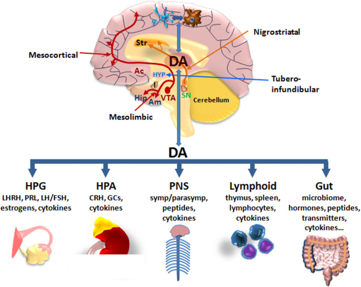
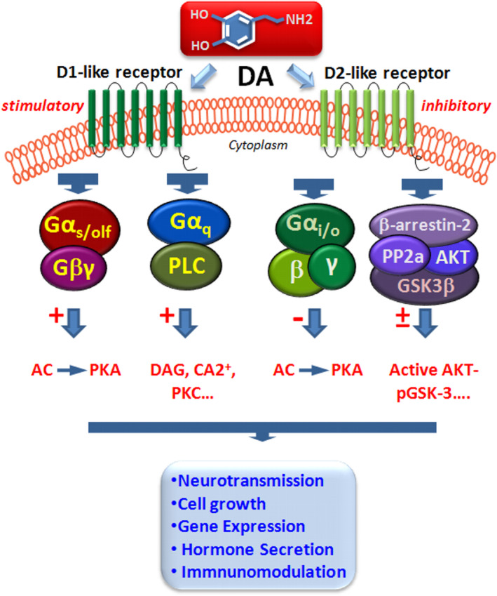
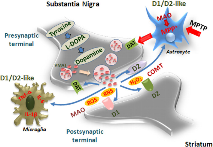
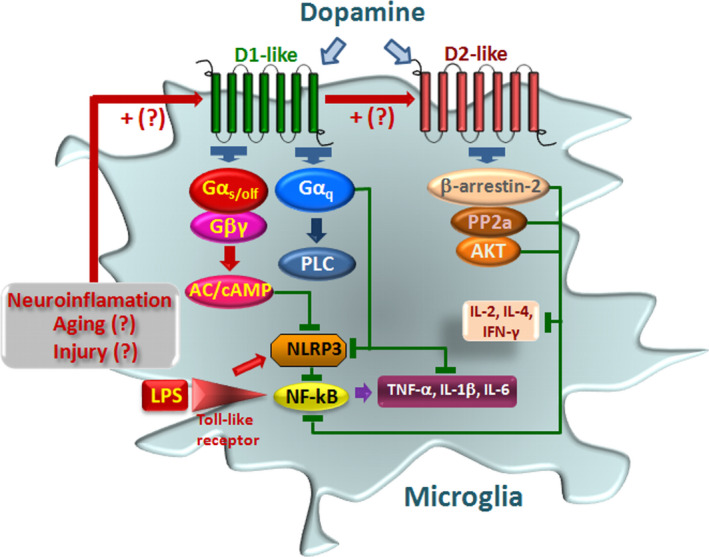
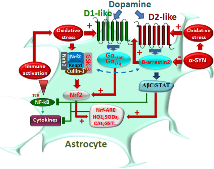
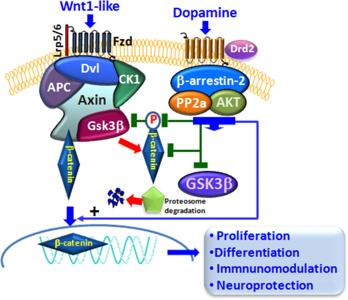
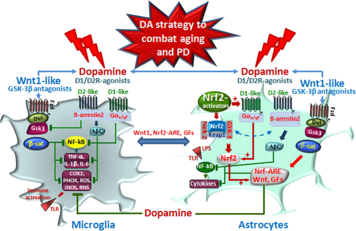
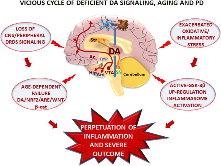
Dopamine (DA) signaling via G protein-coupled receptors is a multifunctional neurotransmitter and neuroendocrine-immune modulator. The DA nigrostriatal pathway, which controls the motor coordination, progressively degenerates in Parkinson's disease (PD), a most common neurodegenerative disorder (ND) characterized by a selective, age-dependent loss of substantia nigra pars compacta (SNpc) neurons, where DA itself is a primary source of oxidative stress and mitochondrial impairment, intersecting astrocyte and microglial inflammatory networks. Importantly, glia acts as a preferential neuroendocrine-immune DA target, in turn, counter-modulating inflammatory processes. With a major focus on DA intersection within the astrocyte-microglial inflammatory network in PD vulnerability, we herein first summarize the characteristics of DA signaling systems, the propensity of DA neurons to oxidative stress, and glial inflammatory triggers dictating the vulnerability to PD. Reciprocally, DA modulation of astrocytes and microglial reactivity, coupled to the synergic impact of gene-environment interactions, then constitute a further level of control regulating midbrain DA neuron (mDAn) survival/death. Not surprisingly, within this circuitry, DA converges to modulate nuclear factor erythroid 2-like 2 (Nrf2), the master regulator of cellular defense against oxidative stress and inflammation, and Wingless (Wnt)/β-catenin signaling, a key pathway for mDAn neurogenesis, neuroprotection, and immunomodulation, adding to the already complex "signaling puzzle," a novel actor in mDAn-glial regulatory machinery. Here, we propose an autoregulatory feedback system allowing DA to act as an endogenous Nrf2/Wnt innate modulator and trace the importance of DA receptor agonists applied to the clinic as immune modifiers.

Keywords: Nrf2/Wnt signaling; Parkinson's disease; dopamine signaling; glial-neuron crosstalk; inflammation; oxidative stress.


Conflict of interest statement

We declare no conflict of interest.

Figures

FIGURE 1
FIGURE 1
Dopamine as a neuroendocrine–immunomodulator. Schematic representation of DA pathways in CNS and bidirectional DA crosstalk at central and peripheral levels orchestrating the regulation of neuroendocrine, autonomic, lymphoid, and gut axes. Bidirectional circuits linking brain DA to astrocyte and microglial crosstalk are schematically represented. There are three major DA pathways in the brain. The nigrostriatal DA pathway originating in the substantia nigra pars compacta (SNpc, A9) releases DA into the corpus striatum (Str), which governs motor coordination. The mesocortical and mesolimbic DA pathways arise from the ventral tegmental area (VTA, A10), releasing DA into major brain limbic regions, including the nucleus accumbens (Ac), the amygdala (Am), the hippocampus (Hip), and the prefrontal cortex, constituting the mesolimbic–mesocortical reward pathway. Within the hypothalamus (HYP), the tuberoinfundibular DA system modulates the output of releasing factors regulating the hypothalamic–hypophyseal–gonadal (HPG) and hypothalamic–hypophyseal–adrenocortical (HPA) axes, neuropeptides, and hormones, including luteinizing hormone‐releasing hormone (LHRH) and prolactin (PRL), in turn involved in immunomodulation. At peripheral level, DA can communicate with the immune system to modulate its activity, directly through specific receptors in immune organs and cells or indirectly through the peripheral nervous system (PNS), via sympathetic and parasympathetic innervation, neuropeptides, and hormone release. Bidirectional DA crosstalk between CNS and gastrointestinal DA, within the brain–gut axis, also plays roles in modulating microenvironmental cues, including the inflammatory milieu and microbiome homeostasis
FIGURE 2
FIGURE 2
Dopamine receptors and signaling pathways in neuroimmune network. Simplified schematic representation of DA acting via DRD1‐ and DRD2‐like receptors by G protein‐dependent, by stimulatory (Gαs) or inhibitory Gαi/o subunits, or by G protein‐independent β‐arrestin‐2 (βArr2)‐dependent pathway (for details, see the text). DA binding to DRD1‐like receptor subtypes can elicit two transduction pathways, of which one is coupled to Gαs/olf, driving adenylyl cyclase increasing cyclic adenosine monophosphate (cAMP) activity. In addition to DRD1 effects on cAMP‐regulated signaling, DRD1Rs couple to Gαq to modulate phospholipase C (PLC) pathway, in turn activating phospholipid turnover and increasing diacylglycerol (DAG), releasing Ca2+ from internal stores, and activating protein kinase C (PKC). D2‐like receptor subtypes, coupled to Gαi/o, suppress cAMP activity, thereby producing an inhibitory effect upon DA binding. The G protein‐independent D2R signaling is represented by βArr2‐mediated signaling. The activation of the D2‐like receptors contributes to the constitution of a protein complex composed of protein phosphatase 2A (PP2A), serine/threonine kinase (Akt), and βArr2, where PP2A increases the dephosphorylation and inactivation of Akt, leading to the modulation of glycogen synthase kinase‐3 (GSK‐3) activation
FIGURE 3
FIGURE 3
Dopamine metabolic pathways and astrocyte–microglial oxidative/inflammatory network. A schematic view of DA pre/postsynaptic regulatory functions. DA biosynthetic steps start with the action of the enzyme tyrosine hydroxylase (TH), the rate‐limiting step in the biosynthesis of DA in the presynaptic terminals to form the DA precursor, L‐DOPA, the principal drug in the therapeutic management of PD. Next, L‐DOPA is decarboxylated to form DA. DA is next incorporated into synaptic vesicles, via the vesicular monoamine transporter 2 (VMAT2), permitting its protection from metabolic inactivation, and its storage until stimulation, when DA released by exocytosis then reaches postsynaptic neurons and binds to cognate D1‐ and D2‐like receptors. D2 presynaptic (inhibitory) receptor can stop the further production and release of DA. The reuptake of DA by presynaptic terminals through the actions of the high‐affinity DA transporter (DT) represents another key step whereby DA is recycled back into the storage vesicles, responsible for modulating the concentration of extraneuronal DA in the brain. Two enzymes are responsible for DA inactivation, monoamine oxidases (MAOs) and catechol‐O‐methyl transferase (COMT), predominantly expressed by astrocytes. During DA metabolic steps, reactive oxygen (ROS) and nitrogen (RNS) species can be produced, which may further engender a neurotoxic cycle capable of causing cell death (for details, see the text). Astrocyte–neuron dialogue may be harmful upon exposure to 1‐methyl‐4‐phenyl‐1,2,3,6‐ tetrahydropyridine (MPTP), as the neurotoxin is converted to its active metabolite in astrocytes, MPP+, then specifically transported by DAT and concentrated within the nigral DA neurons where it inhibits complex I of the mitochondrial electron transport chain, resulting in ATP depletion and subsequent neuronal cell death. This process associated with a robust microgliosis and proinflammatory cytokines, tumor necrosis factor α (TNF‐α), and interleukin‐1β (IL‐1 β) production can be counter‐modulated by DA anti‐inflammatory effects via D1/D2‐like receptors in glial cells, as discussed in Sections 1.3–1.5
FIGURE 4
FIGURE 4
Dopamine signaling pathways modulate inflammasome activation in microglia. Immune activation is schematically represented. LPS via Toll‐like receptors (TLRs) activates Nod‐like receptor protein 3 (NLRP3) inflammasome and nuclear factor kappa‐light‐chain‐enhancer of activated B cell (NF‐ĸB) signaling pathways promoting proinflammatory cytokine (IL‐1β, TNF‐α, IL‐6) release (detailed in Section 1.2). DA and DA agonist activation of D1‐like receptors (D1 and D5) results in a downmodulation of immune response. D1 activation via Gαsolf increases cAMP, which binds directly to NLRP3 triggering its ubiquitination via an autophagy‐mediated degradation. Activated cAMP signaling also inhibits p65/RelA and p50 activation. D5R activation directly recruits a multiprotein complex, impairing activation of NF‐kB. Activation of D2R‐b‐arrestin‐2 complex also results in D2R binding to NLRP3 to repress its activation. D2R signaling can negatively regulate the NF‐kB signaling pathway, thereby inhibiting major proinflammatory cytokine release. The hypothetical role of neuroinflammation, aging, and brain injury, as a counter‐regulatory mechanism, via upregulation of DA receptor expression is illustrated
FIGURE 5
FIGURE 5
Dopamine signaling pathways intersect oxidative/inflammatory cascades in astrocytes. Schematic representation of DA modulation of astrocyte harmful phenotype during inflammation and oxidative stress. DA crosstalk with Nrf2‐ARE induced targeting of antioxidant response elements (ARE) is highlighted. Upon DA binding to DRD2, neuroinflammation can be mitigated by different mechanisms. αB‐Crystallin (αBC)‐dependent mechanism can be elicited by DRD2 agonists alleviating neuroinflammatory injury via the αβC/STAT3 pathway. DRD2 agonists can also mitigate LPS‐induced proinflammatory cytokine response, via a β‐arrestin‐2‐mediated signaling inhibiting NLRP3 inflammasome activation. On the contrary, α‐Syn reduced the expression of β‐arrestin‐2 in astrocytes, whereas it increased the β‐arrestin‐2 and can restore the anti‐inflammatory effect of DRD2 (detailed in the text). A critical loop is represented by the ability of DRD signaling to upregulate the master regulator of oxidative stress and inflammation, Nrf2 in astrocytes, via ARE stimulation of a panel antioxidant/anti‐inflammatory proteins, such as heme oxygenase (HO1), superoxide dismutases (SODs), glutathione S‐transferase (GST), and catalase (CAT) besides others, regulating the cellular redox state by decreasing oxidative stress and inflammation (detailed in Section 1.4)
FIGURE 6
FIGURE 6
Dopamine signaling pathways crosstalk with Wnt/β‐catenin/GSK‐3β cascade. Simplified representation of the Wnt/β‐catenin signaling pathway and its intersection with DRD2 signaling. Wnt signal activation is tightly controlled by a dynamic signaling complex comprised of core receptors from the Frizzled (Fzds) family of G protein‐coupled receptors (GPCRs), the low‐density lipoprotein (LDL) receptor‐related protein (LRP) 5/6 co‐receptors, and the disheveled (Dvl) and Axin adapters. Binding of Wnt1‐like endogenous/exogenous agonists to Fzd triggers a molecular cascade leading to the cytoplasmic accumulation of β‐catenin, which enters the nucleus, and associates with T‐cell factor/lymphoid enhancer binding factor (TCF/LEF) transcription factors, in turn promoting the transcription of Wnt target genes. β‐Catenin is tightly regulated via phosphorylation by the ‘destruction complex’, consisting of glycogen synthase kinase‐3β (GSK‐3β), casein kinase 1α (CK1α), the scaffold protein Axin, and the tumor suppressor adenomatous polyposis coli (APC). DRD2 downstream intracellular G protein‐independent, arrestin‐dependent pathways can target Wnt/β‐catenin signaling, intersecting GSK‐3β, through the contribution serine/threonine kinase (AKT)‐mediated phosphorylation. Crosstalk between DRD2 and Wnt signaling can relieve β‐catenin from active GSK‐3 phosphorylation, thus permitting β‐catenin translocation in the nucleus activating transcription of Wnt‐dependent genes involved in proliferation, differentiation, neuroprotection and immunomodulation (detailed in the text)
FIGURE 7
FIGURE 7
Dopamine drives astrocyte–microglial crosstalk via DRDs/Nrf2/Wnt/GSK‐3 signaling to combat oxidative stress and inflammation. Schematic representation of DA signaling pathways intersecting major oxidative/inflammatory networks in astrocyte–microglial dialogue in PD. Aging, inflammation, and toxic (including bacterial, viral, neurotoxic…) exposures work in synergy with genetic mutations impair nigrostriatal DA neurons. DA and DA agonist can revert such harmful dialogue via a glial switch toward a beneficial antioxidant/anti‐inflammatory and neuroprotective phenotype. DA and DA agonists acting via DRD1 and DRD2 in astrocytes can upregulate Nrf2/HO1 and Wnt1/βcatenin during oxidative stress and inflammation representing a self‐defense system for mDAn survival. Increased DRD2‐β‐arrestin‐2/AKT cascade may then block GSK‐3β‐induced phosphorylation and proteasomal degradation of the neuronal pool of β‐catenin. Stabilized β‐catenin can translocate into the nucleus and associate with a family of transcription factors and regulate the expression of Wnt target genes involved in DA neuron survival/plasticity, neuroprotection and repair. Oxidative stress engendered by DA itself may also function as a critical negative feedback mechanism via DRD5 induction Nrf2‐ARE cascade and/or via DRD2/β‐arrestin‐2‐induced GSK‐3 inhibition, leading to Nrf2 nuclear translocation. DA‐induced beneficial astrocyte phenotype also intersects microglial inflammatory phenotype via both direct DRD1 and DRD2 transduction pathways inhibiting NLRP3/ NF‐ĸB cascade, and/or via astrocyte beneficial feedback onto microglial cells, via astrocytic Wnt1‐like ligands through Fzd receptors, GSK‐3β antagonist, or HO1‐induced anti‐inflammatory effects
FIGURE 8
FIGURE 8
Vicious cycle of dopamine deficiency, aging, inflammation, and CNS disease. Schematic illustration of the interacting harmful cascades arising from DA deficiency at central and peripheral levels engendering a detrimental vicious cycle. The dramatic loss of DA‐mediated signaling at central and peripheral levels associated with the age‐ and PD‐dependent GSK‐3β overactivation in turn creates a favorable milieu driving a feedforward cycle of inflammation/neurodegeneration, as loss of Nrf2/Wnt and upregulation of GSK‐3 phosphorylating and degrading β‐catenin further drive inflammation and excessive oxidative stress associated with inhibition of adult neurogenesis and neurorepair (Marchetti, 2020)

References

    1. Abdalkader, M. , Lampinen, R. , Kanninen, K. M. , Malm, T. M. , & Liddell, J. R. (2018). Targeting Nrf2 to Suppress Ferroptosis and Mitochondrial Dysfunction in Neurodegeneration. Frontiers in Neuroscience, 12, 466. 10.3389/fnins.2018.00466 - DOI - PMC - PubMed
    1. Adams, L. , Franco, M. C. , & Estevez, A. G. (2015). Reactive nitrogen species in cellular signaling. Experimental Biology and Medicine, 240(6), 711–717. 10.1177/1535370215581314 - DOI - PMC - PubMed
    1. Afanas'ev, I. B. (2005). On mechanism of superoxide signaling under physiological and pathophysiological conditions. Medical Hypotheses, 64(1), 127–129. 10.1016/j.mehy.2004.05.009 - DOI - PubMed
    1. Afonina, I. S. , Zhong, Z. , Karin, M. , & Beyaert, R. (2017). Limiting inflammation ‐ The negative regulation of NF‐kB and the NLRP3 inflammasome. In Nature immunology (Vol. 18, Issue 8, pp. 861–869). Nature Publishing Group. 10.1038/ni.3772 - DOI - PubMed
    1. Ahmed, S. , Panda, S. , Kwatra, M. , Sahu, B. , & Naidu, V. G. M. (2021). Pharmacological blocker of NF‐κb and mitochondrial Ros restrict NLRP3 inflammasome activation and rescue dopaminergic neurons in vitro and in vivo. Parkinson’s Disease. 10.21203/rs.3.rs-260478/v1 - DOI - PubMed

Publication types